ABSTRACTS/RESEARCH
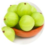
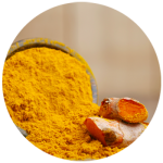
Amla
Indian Gooseberry, also known as “Amla”, is excellent for getting your cholesterol levels into balance. Indian Gooseberry also reduces inflammation one of the primary causes of all disease, especially heart disease and stroke.
-
- A randomized, double blind, placebo controlled, multicenter clinical trial to assess the efficacy and safety of Emblica officinalis extract in patients with dyslipidemia. Background-Dyslipidemia is one of the most frequently implicated risk factors for development of atherosclerosis. This study evaluated the efficacy of amla (Emblica officinalis) extract (composed of polyphenols, triterpenoids, oils etc. as found in the fresh wild amla fruit) in patients with dyslipidemia. Methods-A total of 98 dyslipidemic patients were enrolled and divided into amla and placebo groups. Amla extract (500 mg) or a matching placebo capsule was administered twice daily for 12 weeks to the respective group of patients. The patients were followed up for 12 weeks and efficacy of study medication was assessed by analyzing lipid profile. Other parameters evaluated were apolipoprotein B (Apo B), apolipoprotein A1 (Apo A1), Coenzyme Q10 (CoQ10), high-sensitive C-reactive protein (hsCRP), fasting blood sugar (FBS), homocysteine and thyroid stimulating hormone (TSH). Results-In 12 weeks, the major lipids such as total cholesterol (TC) (p = 0.0003), triglyceride (TG) (p = 0.0003), low density lipoprotein cholesterol (LDL-C) (p = 0.0064) and very low density lipoprotein cholesterol (VLDL-C) (p = 0.0001) were significantly lower in amla group as compared to placebo group. Additionally, a 39% reduction in atherogenic index of the plasma (AIP) (p = 0.0177) was also noted in amla group. The ratio of Apo B to Apo A1 was reduced more (p = 0.0866) in the amla group as compared to the placebo. There was no significant change in CoQ10 level of amla (p = 0.2942) or placebo groups (p = 0.6744). Although there was a general trend of FBS reduction, the numbers of participants who may be classified as pre-diabetes and diabetes groups (FBS > 100 mg/dl) in the amla group were only 8. These results show that the amla extract used in the study is potentially a hypoglycaemic as well. However, this needs reconfirmation in a larger study. Conclusions-The Amla extract has shown significant potential in reducing TC and TG levels as well as lipid ratios, AIP and apoB/apo A-I in dyslipidemic persons and thus has scope to treat general as well as diabetic dyslipidemia. A single agent to reduce cholesterol as well as TG is rare. Cholesterol reduction is achieved without concomitant reduction of Co Q10, in contrast to what is observed with statins.[ Haridas Upadya, S. Prabhu, Aravinda Prasad, Deepa Subramanian, Swati Gupta & Ajay Goel BMC Complementary and Alternative Medicine volume 19, Article number: 27 (2019)]
- Amlamax in the Management of Dyslipidemia in Humans. Hypercholesterolemia is the major cause of cardiovascular diseases leading to myocardial infractions leading to considerable morbidity and mortality. During the past decade a group of molecules referred to as statins such as simvastatin, atrovastatin have been tried with great success in reducing total cholesterol. These molecules act by inhibiting the HMG CoA reductase enzyme thereby interfering with the synthesis of cholesterol. But statins reduce all the cholesterol including HDL cholesterol. Long term drug vigilance activity has revealed serious side effects of tendinopathy and related musculoskeletal disorders in some of the subjects. In an effort to manage hypercholesterolemia without serious side effects in a natural way we had tried the use of Amlamax a reconstituted, purified, standardized dried extract of amla (Emblica officinalis) containing 30% ellagitannins significant elevation of HDL cholesterol by the administration of Amlamax [Antony B, Merina B, Sheeba, V. Amlamax in the Management of Dyslipidemia in Humans. Indian J Pharm Sci. 2008 Jul-Aug; 70(4): 504-507]
- A Pilot Clinical Study to Evaluate the Effect of Emblica Officinalis Extract (Amlamax) on Markers of Systemic Inflammation and Dyslipidemia. Emblica officinalis Gaertn, commonly known as the Indian gooseberry of “Amla”, has been used as health food for centuries in India and other Asian countries. The biological effects of amla have been attributed to the antioxidant properties of the low-molecular weight hydrolysable tannins present in the fruit. Amlamax is a purified, standardized, dried extract of amla containing about 35% galloellagi tannins along with other hydrolysable tannins. Our earlier studies on rabbits showed significant reduction in total cholesterol and triglycerides as well as increase in HDL. The present study extends these results to human volunteers. Two doses of the extract were evaluated – 500mg and 1000mg per day for 6 months. Blood samples were collected at the 3rd and 6th months showed reduction in total and LDL cholesterols and enhancement of beneficial HDL cholesterol. In addition, blood CRP levels, a marker for inflammation, were also significantly reduced. Since dyslipidemia and inflammation and the two major components of cardiovascular diseases, the present results must be considered encouraging and indicate the potential of Amlamax in the management of heart disease. [Antony B, Benny M, KaimalTNB. A Pilot Clinical Study to Evaluate the Effect of Emblica Officinalis Extract (Amlamax) on Markers of Systemic Inflammation and Dyslipidemia. Indian Journal of Clinical Biochemistry. 2008; 23(4): 378-381.]
- Effect of Standardized Amla Extract on Atherosclerosis and Dyslipidemia. Emblica officinalis, commonly known as Indian gooseberry (Amla), is found to be effective for the reversal of dyslipidemia and intima-media thickening and plaque formation in the aorta in hypercholesterolaemic rabbits. In this study, cholesterol powder (100 mg/kg body weight) was administered orally to healthy NZ white rabbits for 4 mo to induce hypercholesterolaemia; and thereafter, amla extract was given in two doses (10 mg and 20 mg/kg/ d orally) for 4 mo. Fasting lipid profile was done monthly and also at the end of treatment. After sacrificing the animals, tissue cholesterol (liver, heart and kidney) and 3-hydroxy-3-methylglutaryl-Coenzyme A reductase activity of liver were estimated and part of aorta and myocardium were processed for histological studies. Feeding of amla extract (10 mg and 20 mg/kg) for 4 mo reversed these changes and the lumen of the aorta became normal as in the normal control group. Reversal of dyslipidemia and atheromatous plaques achieved by amla extract seems to be brought about by a number of factors, such as its ability to prevent low-density lipoprotein oxidation, its antioxidant action, besides decreasing synthesis of cholesterol by inhibiting 3-hydroxy-3-methylglutaryl-Coenzyme A reductase activity and elevating high-density lipoprotein level to enhance reverse cholesterol transport. [Antony B, Merina B, Sheeba V, Mukkadan J. Effect of Standardized Amla Extract on Atherosclerosis and Dyslipidemia. Indian J Pharm Sci. July-August 2006; 68(4): 437-441.]
- Toxicity Studies of Amlamax – Purified Standardized Extract of Emblica Officinalis. Amlamax is the purified, standardized extract of ‘Amla’ fruits. Acute and subacute toxicity studies were carried out by oral administration of Amlamax at different dosages in mice and rats. Doses up to 2g/kg orally for three months did not produce any haematological, biochemical and histological changes in vital organs.[Antony B, Merina B, Sheeba V. Toxicity Studies of Amlamax – Purified Standardized Extract of Emblica Officinalis. Indian J. Nat. Prod. 2007;23(2):14-17.]
- Hypolipidemic Activity of Phyllanthus Emblica Linn (Amla) & Trigonella Foenum Graecum (Fenugreenk) Combination In Hypercholesterolemic Subjects – A Prospective, Randomised, Parallel, Open-Label, Positive Controlled Study. The present study is aimed at to evaluate the hypolipidemic and antioxidant activity of combination of fenugreek and amla in hyperlipidemic subjects.Twenty eight subjects were selected from outpatient department of medicine with the inclusion criteria of total cholesterol > 220mg/dl and or serum triglyceride >140mg/dl. They were enrolled for a period of 12 weeks after obtaining written informed consent. Subjects were randomly divided into 2 groups. Group I [test drug combination -1000mg Amla + 500mg Fenugreek/day] and Group II [standard drug Atorvastatin 10 mg daily]. Subjects were evaluated every 4 weeks for 12 weeks. During these visits biochemical investigations of lipid profile and antioxidant activity were done. At the end of 12 weeks, there were significant decrease in serum total cholesterol (20-26%), LDL (25-34%), triglyceride (15-30%) and VLDL (15-30%) levels & an increase in the HDL (0-5%) in the test drug group. This combination has shown similar response on lipid profile as statins with lesser adverse effects. Antioxidant activity of the combination is also demonstrated in the trial. [Joseph S, Santhost D, Udupa AL, Gupta S, Ojeh N, Rathnakar UP, et al. Hypolipidemic Activity of Phyllanthus Emblica Linn (Amla) & Trigonella Foenum Graecum (Fenugreek) Combination In Hypercholesterolemic Subjects – A Prospective, Randomised, Parallel, Open-Label, Positive Controlled Study. Asian Journal of Biochemical and Pharmaceutical Research. 2012;1(2):225-230.]
Read More..
Read Less..
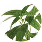
Andrographis
Andrographis paniculata, an adaptogenic herb used in traditional Ayurvedic practice, can be a powerful ally in the fight against cancer.
- Curcumin and andrographis exhibit anti-tumor effects in colorectal cancer via activation of ferroptosis and dual suppression of glutathione peroxidase-4 and ferroptosis suppressor protein-1. Abstract: Colorectal cancer (CRC) is the leading cause of cancer-related deaths worldwide. The limitations of current chemotherapeutic drugs in CRC include their toxicity, side effects, and exorbitant costs. To assess these unmet needs in CRC treatment, several naturally occurring compounds, including curcumin and andrographis, have gained increasing attention due to their multi-targeted functionality and safety vs. conventional drugs. In the current study, we revealed that a combination of curcumin and andrographis exhibited superior anti-tumor effects by inhibiting cell proliferation, invasion, colony formation, and inducing apoptosis. Genome-wide transcriptomic expression profiling analysis revealed that curcumin and andrographis activated the ferroptosis pathway. Moreover, we confirmed the gene and protein expression of glutathione peroxidase 4 (GPX-4) and ferroptosis suppressor protein 1 (FSP-1), the two major negative regulators of ferroptosis, were downregulated by this combined treatment. With this regimen, we also observed that intracellular accumulation of reactive oxygen species and lipid peroxides were induced in CRC cells. These cell line findings were validated in patient-derived organoids. In conclusion, our study revealed that combined treatment with curcumin and andrographis exhibited anti-tumorigenic effects in CRC cells through activation of ferroptosis and by dual suppression of GPX-4 and FSP-1, which have significant potential implications for the adjunctive treatment of CRC patients. [Miyazaki K, Xu C, Shimada M, Goel A. Curcumin and andrographis exhibit anti-tumor effects in colorectal cancer via activation of ferroptosis and dual suppression of glutathione peroxidase-4 and ferroptosis suppressor protein-1. Pharmaceuticals. 2023; 16:383.]
- Andrographis reverses gemcitabine resistance through regulation of ERBB3 and calcium signaling pathway in pancreatic ductal adenocarcinoma. Pancreatic ductal adenocarcinoma (PDAC) is one of the most lethal malignancies, primarily due to intrinsic or acquired resistance to chemotherapy, such as Gemcitabine (Gem). Naturally occurring botanicals, including Andrographis (Andro), can help enhance the anti-tumorigenic therapeutic efficacy of conventional chemotherapy through time-tested safety and cost effectiveness. Accordingly, we hypothesized that Andro might reverse Gem resistance in PDAC. The critical regulatory pathways associated with Gem resistance in PDAC were identified by analyzing publicly available transcriptomic profiling and PDAC tissue specimens. A series of systematic in vitro experiments were performed using Gem-resistant (Gem-R) PDAC cells and patient derived 3D-organoids to evaluate the Andro-mediated reversal of Gem resistance in PDAC. Transcriptomic profiling identified the calcium signaling pathway as a critical regulator of Gem-resistance (Fold enrichment: 2.8, p = 0.002). Within this pathway, high ERBB3 expression was significantly associated with poor prognosis in PDAC patients. The combination of Andro and Gem exhibited superior anti-cancer potential in Gem-R PDAC cells through potentiating cellular apoptosis. The combined treatment down-regulated ERBB3 and decreased intracellular calcium concentration in Gem-R PDAC cells. Finally, these findings were successfully interrogated in patient-derived 3D-organoids. In conclusion, we demonstrate novel evidence for Andro-mediated reversal of chemoresistance to Gem in PDAC cells through the regulation of ERBB3 and calcium signaling. [Okuno K, Xu C, Pascual-Sabater S, et al. Andrographis reverses gemcitabine resistance through regulation of ERBB3 and calcium signaling pathway in pancreatic ductal adenocarcinoma. Biomedicines. 2023;11:119.]
- A combined treatment with berberine and andrographis exhibits enhanced anti-cancer activity through suppression of DNA replication in colorectal cancer.
The high morbidity and mortality associated with colorectal cancer (CRC) are largely due to the invariable development of chemoresistance to classic chemotherapies, as well as intolerance to their significant toxicity. Many pharmaceutical formulations screened from natural plant extracts offer safe, inexpensive, and multi-target therapeutic options. In this study, we demonstrated that Berberis vulgaris L. (Berberine) and Andrographis paniculata (Burm. f.) Nees (Andrographis) extracts exerted their synergistic amplified anti-cancer effects by jointly inhibiting cell viability, suppressing colony formation, and inducing cell cycle arrest. Consistent with our in-vitro findings, the amplified synergistic anti-cancer effects were also observed in subcutaneous xenograft preclinical animal models, as well as patient-derived primary tumor organoids. To explore the molecular mechanisms underlying the amplified synergistic anti-cancer effects, RNA sequencing was performed to identify candidate pathways and genes. A transcriptome analysis revealed that DNA-replication-related genes, including FEN1, MCM7, PRIM1, MCM5, POLA1, MCM4, and PCNA, may be responsible for the enhanced anticancer effects of these two natural extracts. Taken together, our data revealed the powerful enhanced synergistic anti-CRC effects of berberine and Andrographis and provide evidence for the combinational targeting of DNA-replication-related genes as a promising new strategy for the therapeutic option in the management of CRC patients. [Zhao Y, Roy S, Want C, Goel A. A combined treatment with berberine and andrographis exhibits enhanced anti-cancer activity through suppression of DNA replication in colorectal cancer. Pharmaceuticals.2022;15:262.] - A combined treatment with melatonin and andrographis promotes autophagy and anti-cancer activity in colorectal cancer.
Colorectal cancer (CRC) is one of the most frequent malignancies worldwide and remains one of the leading causes of cancer-related deaths in the United States. The high degree of morbidity and mortality associated with this disease is largely due to the inadequate efficacy of current treatments as well the development of chemoresistance. In recent years, several pharmaceutical agents screened from natural products have shown the promise to offer a safe, inexpensive, and synergistically multi-targeted treatment option in various cancer. Given the growing evidence of anti-carcinogenic properties of two natural compounds, melatonin (MLT) and andrographis (Andro), we aimed to evaluate their synergistic anti-cancer effects in CRC. We demonstrate that indeed these two compounds possessed a synergistic anti-cancer effect in terms of their ability to inhibit cell viability, suppression of colony-formation and induction of apoptosis (p<0.05). In line with our in-vitro findings, we were able to validate this combinatorial anti-cancer activity in xenograft animal models (p<0.001) as well as tumor-derived 3D organoids (p<0.01). RNA-sequencing analysis revealed candidate pathways and genes that mediated anti-tumor efficacy of MLT and Andro in CRC, among which autophagy pathway and related genes, including NR4A1, CTSL and Atg12, were found to be primarily responsible for the increased anti-cancer effect by the two natural products. In conclusion, our data reveal a potent and synergistic therapeutic effect of MLT and Andro in the treatment of CRC and provides a rationale for suppressing autophagy in cancer cells as a potential therapeutic strategy for CRC. [Zhao Y, Wang C, Goel A. A combined treatment with melatonin and andrographis promotes autophagy and anti-cancer activity in colorectal cancer. Carcinogenesis. 2022; January. Published online ahead of print. - Andrographis-mediated chemosensitization through activation of ferroptosis and suppression of β-catenin/Wnt-signaling pathways in colorectal cancer.
Colorectal cancer (CRC) remains one of the leading causes of cancer-related mortality in the USA. As much as 50–60% of CRC patients develop resistance to 5-fluorouracil (5FU)-based chemotherapeutic regimens, attributing the increased overall morbidity and mortality. In view of the growing evidence that active principles in various naturally occurring botanicals can facilitate chemosensitization in cancer cells, herein, we undertook a comprehensive effort in interrogating the activity of one such botanical—andrographis—by analyzing its activity in CRC cell lines [both sensitive and 5FU resistant (5FUR)], a xenograft animal model and patient-derived tumor organoids. We observed that combined treatment with andrographis was synergistic and resulted in a significant and dose-dependent increase in the efficacy of 5FU in HCT116 and SW480 5FUR cells (P < 0.05), reduced clonogenic formation (P < 0.01) and increased rates of caspase-9-mediated apoptosis (P < 0.05). The genomewide expression analysis in cell lines led us to uncover that activation of ferroptosis and suppression of β-catenin/Wnt-signaling pathways were the key mediators for the anti-cancer and chemosensitizing effects of andrographis. Subsequently, we validated our findings in a xenograft animal model, as well as two independent CRC patient-derived organoids—which confirmed that combined treatment with Andrographis was significantly more effective than 5FU and andrographis alone and that these effects were in part orchestrated through dysregulated expression of key genes (including HMOX1, GCLC, GCLM and TCF7L2) within the ferroptosis and Wnt-signaling pathways. Collectively, our data highlight that andrographis might offer a safe and inexpensive adjunctive therapeutic option in the management of CRC patients. [Sharma P, Shimura T, Banwait JK, Goel A. Andrographis-mediated chemosensitization through activation of ferroptosis and suppression of β-catenin/Wnt-signaling pathways in colorectal cancer. Carcinogenesis. 2020:1-10. - Andrographis overcomes 5-fluorouracil associated chemoresistance through inhibition of DKK1 in colorectal cancer.
Colorectal cancer (CRC) ranks as the third leading cause of cancer-related deaths in the US. 5-fluorouracil (5FU)-based chemotherapeutic drug remains a mainstay of CRC treatment. Unfortunately, ~50-60% of patients eventually develop resistance to 5FU, leading to poor survival outcomes. Our previous work revealed that andrographis enhanced 5FU-induced anti-cancer activity, but the underlying mechanistic understanding largely remains unclear. In this study, we first established 5FU resistant (5FUR) CRC cells and observed that combined treatment with andrographis-5FU in 5FUR cells exhibited superior effect on cell viability, proliferation and colony formation capacity compared to individual treatments (p<0.001). To identify key genes and pathways responsible for 5FU resistance, we analyzed genome-wide transcriptomic profiling data from CRC patients who either responded or did not respond to 5FU. Among a panel of differentially expressed genes, DKK1 overexpression was a critical event for 5FU resistance. Moreover, andrographis significantly downregulated 5FU-induced DKK1 overexpression, accompanied with enhanced anti-tumor effects by abrogating downstream Akt-phosphorylation. In line with in vitro findings, andrographis enhanced 5FU-induced anti-cancer activity in mice xenografts and patient-derived tumoroids (p<0.01). In conclusion, our data provide novel evidence for andrographis-mediated reversal of 5FU resistance, highlighting its potential role as an adjunct to conventional chemotherapy in CRC. [Zhao Y, Wang C, Goel A. Andrographis overcomes 5-fluorouracil associated chemoresistance through inhibition of DKK1 in colorectal cancer. Carcinogenesis. 2021. Advanced publication data: https://doi.org/10.1093/carcin/bgab027] - Enhanced anti-cancer activity of andrographis with oligomeric proanthocyanidins through activation of metabolic and ferroptosis pathways in colorectal cancer.
The high degree of morbidity and mortality in colorectal cancer (CRC) patients is largely due to the development of chemoresistance against conventional chemotherapeutic drugs. In view of the accumulating evidence that various dietary botanicals ofer a safe, inexpensive and multi-targeted treatment option, herein, we hypothesized that a combination of Andrographis paniculata and Oligomeric Proanthocyanidins (OPCs) might interact together with regard to anti-tumorigenic activity in CRC. As a result, we demonstrated the enhanced anti-cancer activity between these two botanical extracts in terms of their ability to inhibit cancer cell growth, suppress colony formation and induce apoptosis. Furthermore, we validated these fndings in subcutaneous xenograft model and in patient derived primary epithelial 3D organoids. Transcriptomic profling identifed involvement of metabolic pathways and ferroptosis-associated genes, including HMOX1, GCLC and GCLM, that may be responsible for the increased anti-tumorigenic activity by the two compounds. Collectively, our study provides novel evidence in support of the combinatorial use of andrographis and OPCs as a potential therapeutic option, perhaps as an adjunctive treatment to classical drugs, in patients with colorectal cancer. [Shimura T, Sharma P, Sharma GG, Banwait JK, Goel A. Enhanced anti-cancer activity of andrographis with oligomeric proanthocyanidins through activation of metabolic and ferroptosis pathways in colorectal cancer. Sci Rep. 2021;11(1):7548.] - Antitumor effects of Andrographis via ferroptosis-associated genes in gastric cancer.
The overall prognosis of advanced/metastatic gastric cancer (GC) remains poor despite the development of pharmacotherapy. Therefore, other treatment options, such as complementary and alternative medicine, should be considered to overcome this aggressive malignancy. Andrographis, which is a generally unharmful botanical compound, has gained increasing interest for its anticancer effects in multiple malignancies via the regulation of cancer progression-associated signaling pathways. In the present study, a series of in vitro experiments (cell proliferation, colony formation and apoptosis assays) was designed to elucidate the antitumor potential and mechanism of Andrographis in GC cells. The present study demonstrated that Andrographis exerted antitumor effects in GC cell lines (MKN74 and NUGC4) by inhibiting proliferation, reducing colony formation and enhancing apoptotic activity. Furthermore, it was demonstrated that the expression levels of the ferroptosis-associated genes heme oxygenase-1, glutamate-cysteine ligase catalytic and glutamate-cysteine ligase modifier were significantly upregulated after Andrographis treatment in both GC cell lines in reverse transcription-quantitative PCR experiments (P<0.05); this finding was further confirmed by immunoblotting assays (P<0.05). In conclusion, to the best of our knowledge, the present study was the first to demonstrate that Andrographis possessed antitumor properties by altering the expression levels of ferroptosis-associated genes, thereby providing novel insights into the potential of Andrographis as an adjunctive treatment option for patients with metastatic GC. [Ma R, Shimura T, Yin C, et al. Antitumor effects of andrographis via ferroptosis-associated genes in gastric cancer. Oncol Lett. 2021;22(1):523.]
Read More..
Read Less..
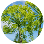
Angelica
Angelica archangelica works directly on the bladder making it the perfect choice for men and women. This amazing extract comes from angelica (Angelica archangelica), which grows in Iceland. This extract comes from the leaves.
-
- A paralled, randomized, double-blind, placebo-controlled study to investigate the effect of Angelica archangelica on nocturia in men. Objective. This study aimed to investigate the effect of Angelica archangelica leaf, on nocturia. Material and methods. Sixty-nine male patients 45 years or older with at least two nocturnal voids were randomized to receive Angelica archangelica or placebo in a double-blind design for 8 weeks. Voiding diaries were assessed before and after the treatment. Results. The results indicate that Angelica archangelica is safe. The actual number of nocturnal voids (ANV), nocturnal polyuria index (NPi) and nocturnal bladder capacity index (NBC index) decreased in the test population, but there was no significant difference between the treatment groups. Subsequent subgroup analysis showed that Angelica archangelica significantly reduced the NBC index and nocturnal voids per sleeping hour in comparison to the placebo in participants with baseline NBC index above 1.3. When participants with sleep disorders were excluded from this group, ANV was also significantly reduced for the Angelica archangelica group in comparison to the placebo group. Conclusion. Angelica archangelica , made from an extract of the medicinal herb Angelica archangelica, is safe. This study did not show that Angelica archangelica improved nocturia overall compared to placebo. Subgroup analysis suggested a beneficial effect in individuals with decreased nocturnal bladder capacity, which warrants further study. [Sigurdsson S, Geirsson G, Gudmundsdottir H, Egilsdottir PB, Gudbjarnason S. A parallel, randomized, double-blind, placebo-controlled study to investigate the effect of Angelica archangelica on nocturia in men. Scand J Urol. 2013 Feb;47(1):26-32.]
- Antitumour activity of Angelica archangelica leaf extract. Background: The purpose of this study was to examine the effect of a leaf extract from A. archangelica on the growth of Crl mouse breast cancer cells in vitro and in vivo. Materials and Methods: The antiproliferative activity of the extract was measured by 3H-thymidine uptake in the Crl cells in vitro. Twenty mice were injected with the Crl cells, and 11 of them were fed A. archangelica leaf extract, and the progress of the tumours was followed. Results: The leaf extract was mildly antiproliferative on the Crl cells with an EC50 of 87.6 Ìg/ml. The antitumour activity of the extract was expressed in the mice by marked reduction in tumour growth. In the experimental animals, 9 out of 11 mice developed no or very small tumours, whereas control animals, not receiving the extract, developed significantly larger tumours (p<0.01), as estimated by Mann-Whitney U-test. The antitumour activity of the leaf extract could not be explained by the antiproliferative activity of furanocoumarins present in the extract. Conclusion: The results demonstrate the antiproliferative activity in vitro and antitumour activity in vivo of a leaf extract from A. archangelica. [Sigurdsson S, Ogmundsdottir HM, Hallgrimsson J, Gudbjarnason S. Antitumour activity of Angelica archangelica leaf extract. in vivo. 2005;19:191-194.]
Read More..
Read Less..
Berberine
Berberine is an alkaloid compound found in many traditionally used medicinal plants, including Indian Barberry (Berberis aristata) and more recently as a synthesized supplement.
- Berberine Overcomes Gemcitabine-Associated Chemoresistance through Regulation of Rap1/PI3K-Akt Signaling in Pancreatic Ductal Adenocarcinoma.
Background: Gemcitabine (Gem)-based chemotherapy is one of the first-line treatments for pancreatic ductal adenocarcinoma (PDAC). However, its clinical effect is limited due to development of chemoresistance. Various naturally occurring compounds, including Berberine (BBR), provide an anti-cancer efficacy with time-tested safety, individually and in combination with chemotherapeutic drugs.
Methods: Accordingly, we hypothesized that BBR might enhance the chemosensitivity to Gem in PDAC. In this study, cell culture studies using MIA PaCa-2 and BxPC-3 cells, followed by analysis in patient-derived organoids were performed to evaluate the anti-cancer effects of BBR in PDAC. Considering that cancer is a significant manifestation of increased chronic inflammatory stress, systems biology approaches are prudent for the identification of molecular pathways and networks responsible for phytochemical-induced anti-cancer activity, we used these approaches for BBR-mediated chemosensitization to Gem.
Results: Firstly, Gem-resistant (Gem-R) PDAC cells were established, and the combination of BBR and Gem revealed superior anti-cancer efficacy in Gem-R cells. Furthermore, the combination treatment induced cell cycle arrest and apoptosis in Gem-R PDAC cells. Transcriptomic profiling investigated the Rap1 and PI3K-Akt signaling pathway as a key regulator of Gem-resistance and was a key mediator for BBR-mediated chemosensitization in PDAC cells. All cell culture-based findings were successfully validated in patient-derived organoids.
Conclusion: In conclusion, we demonstrate that BBR-mediated reversal of chemoresistance to Gem manifests through Rap1/PI3K-Akt signaling in PDAC. [Okuno K, Xu C, Pascual-Sabater S, Tokunaga M, Han H, Fillat C, Kinugasa Y, Goel A. Pharmaceuticals (Basel). 2022 Sep 28;15(10):1199.]
-
Berberine and oligomeric proanthocyanidins exhibit synergistic efficacy through regulation of PI3K-Akt signaling pathway in colorectal cancer.
Background: Naturally occurring dietary botanicals offer time-tested safety and anti-cancer efficacy, and a combination of certain compounds has shown to overcome the elusive chemotherapeutic resistance, which is of great significance for improving the mortality of patients with colorectal cancer (CRC). Accordingly, herein, we hypothesized that berberine (BBR) and oligomeric proanthocyanidins (OPCs) might regulate synergistically multiple oncogenic pathways to exert a superior anti-cancer activity in CRC.
Methods: We performed a series of cell culture studies, followed by their interrogation in patient-derived organoids to evaluate the synergistic effect of BBR and OPCs against CRC. In addition, by performing whole genome transcriptomic profiling we identified the key targeted genes and pathways regulated by the combined treatment.
Results: We first demonstrated that OPCs facilitated enhanced cellular uptake of BBR in CRC cells by measuring the fluorescent signal of BBR in cells treated individually or their combination. The synergism between BBR and OPCs were investigated in terms of their anti-tumorigenic effect on cell viability, clonogenicity, migration, and invasion. Furthermore, the combination treatment potentiated the cellular apoptosis in an Annexin V binding assay.
Transcriptomic profiling identified oncogene MYB in PI3K-AKT signaling pathway might be critically involved in the anti-tumorigenic properties of the combined treatment. Finally, we successfully validated these findings in patient-derived CRC tumor organoids.
Conclusions: Collectively, we for the first time demonstrate that a combined treatment of BBR and OPCs synergistically promote the anti-tumorigenic properties in CRC possibly through the regulation of cellular apoptosis and oncogene MYB in the PI3K-Akt signaling pathway. [Okunu K, Garg R, Yuan YC, Tokunaga M, Kinugasa Y, Goel A. Berberine and oligomeric proanthocyanidins exhibit synergistic efficacy through regulation of PI3K-Akt signaling pathway in colorectal cancer. Front Oncol. 2022;12:855860]
Read More..
Read Less..

Boswellia
This botanical prevents tumors, fights pain, protects the cardiovascular system, and stops digestive diseases.
-
- Effects of anti-inflammatory and adaptogenic herbal extracts on gene expression of eicosanoids signaling pathways in isolated brain cell. Introduction: The adaptogens modulate expression of genes playing key roles in development of aging-related disorders, which are considered as low-grade systemic inflammatory conditions characterized by an imbalance between pro-and anti-inflammatory eicosanoids.Aim of the Study: We compared the effects of anti-inflammatory and adaptogenic plant extracts on the expression of genes involved in biosynthesis of eicosanoids with the purpose to find those plants, which selectively upregulated the expression of anti-inflammatory lipoxins signaling pathways and inhibited pro-inflammatory signaling pathways associated with biosynthesis of leukotrienes, prostaglandins and thromboxanes. Materials and Methods: We conducted transcriptome-wide RNA sequencing to profile gene expression alterations in T98G neuroglia cells upon treatment of plant extract and analyzed the relevance of deregulated genes to eicosanoids signaling pathways using in silico models. Results: For the first time, we demonstrated that Rhodiola rosea, Withania somnifera and Eleutherococcus senticosus downregulate the expression of key genes (ALOX5AP, DPEP2, LTC4S) involved biosynthesis of leukotrienes A, B, C, D and E, resulting in inhibition of leukotriene signaling pathway suggesting their potential benefits in Alzheimer disease. The common feature for all tested anti-inflammatory plants extracts was related to downregulation of ALOX12, which was also associated with neuroprotective action of these medicinal plants as well as their potential benefits in neurodegenerative diseases. None of tested anti-inflammatory and adaptogenic plants selectively activated the ALOX15-mediated signaling pathway, which is associated with generation anti-inflammatory lipoxins. Almost all tested plants upregulated the expression of the prostaglandin E receptor 3 gene(PTGER3) suggesting their potential benefits in the treatment of cancer. Conclusion: Every single plant tested in this study revealed a specific “signature” on eicosanoid signaling-related gene expression, regardless of their common features as anti-inflammatory or adaptogenic activity. Further studies of the combination of Rhodiola with Withania (Adaptra) for the treatment of Alzheimer disease are required. [Panossian,A, Seo EJ, Efferth T. Effects of anti-inflammatory and adaptogenic herbal extracts on gene expression of eicosanoids signaling pathways in isolated brain cells. Phytomedicine 60 March 2019; https://doi.org/10.1016/j.phymed.2019.152881]
- Curcumin downregulates expression of opioid-related nociception receptor gene (OPRL1) in isolated neuroglia cells. Background: Curcumin (CC) exerts polyvalent pharmacological actions and multi-target effects, including pain relief and anti-nociceptive activity. In combination with Boswellia serrata extract (BS), curcumin shows greater efficacy in knee osteoarthritis management, presumably due to synergistic interaction of the ingredients. Aim: To elucidate the molecular mechanisms underlying the analgesic activity of curcumin and its synergistic interaction with BS. Methods: We performed gene expression profiling by transcriptome-wide mRNA sequencing in human T98G neuroglia cells treated with CC (Curamed®), BS, and the combination of CC and BS (CC-BS; Curamin®), followed by interactive pathways analysis of the regulated genes. Results: Treatment with CC and with CC-BS selectively downregulated opioid-related nociceptin receptor 1 gene (OPRL1) expression by 5.9-fold and 7.2-fold, respectively. No changes were detected in the other canonical opioid receptor genes: OPRK1, OPRD1, and OPRM1. Nociceptin reportedly increases the sensation of pain in supra-spinal pain transduction pathways. Thus, CC and CC-BS may downregulate OPRL1, consequently inhibiting production of the nociception receptor NOP, leading to pain relief. In neuroglia cells, CC and CC-BS inhibited signaling pathways related to opioids, neuropathic pain, neuroinflammation, osteoarthritis, and rheumatoid diseases. CC and CC-BS also downregulated ADAM metallopeptidase gene ADAMTS5 expression by 11.2-fold and 13.5-fold, respectively. ADAMTS5 encodes a peptidase that plays a crucial role in osteoarthritis development via inhibition of a corresponding signaling pathway. Conclusion: Here, we report for the first time that CC and CC-BS act as nociceptin receptor antagonists, selectively downregulating opioid-related nociceptin receptor 1 gene (OPRL1) expression, which is associated with pain relief. BS alone did not affect OPRL1 expression, but rather appears to potentiate the effects of CC via multiple mechanisms, including synergistic interactions of molecular networks. [Seo EJ, Efferth T, Panossian A. Curcumin downregulates expression of opioid-related nociception receptor gene (OPRL1) in isolated neuroglia cells. Phytomedicine. 20 September 2018; https://doi.org/10.1016/j.phymed.2018.090.202]
- Novel molecular mechanisms for the adaptogenic effects of herbal extracts on isolated brain cells using systems biology. Introduction: Adaptogens are natural compounds or plant extracts that increase adaptability and survival of organisms under stress. Adaptogens stimulate cellular and organismal defense systems by activating intracellular and extracellular signaling pathways and expression of stress-activated proteins and neuropeptides. The effects adaptogens on mediators of adaptive stress response and longevity signaling pathways have been reported, but their stress-protective mechanisms are still not fully understood. Aim of the Study: The aim of this study was to identify key molecular mechanisms of adaptogenic plants traditionally used to treat stress and aging-related disorders, i.e., Rhodiola rosea, Eleutherococcus senticosus, Withania somnifera, Rhaponticum carthamoides, and Bryonia alba. Materials and Methods: To investigate the underlying molecular mechanisms of adaptogens, we conducted RNA sequencing to profile gene expression alterations in T98G neuroglia cells upon treatment of adaptogens and analyzed the relevance of deregulated genes to adaptive stress-response signaling pathways using in silico pathway analysis software. Results and Discussion: At least 88 of the 3516 genes regulated by adaptogens were closely associated with adaptive stress response and adaptive stress-response signaling pathways (ASRSPs), including neuronal signaling related to corticotropin-releasing hormone, cAMP-mediated, protein kinase A, and CREB; pathways related to signaling involving CXCR4, melatonin, nitric oxide synthase, GP6, G?s, MAPK, neuroinflammation, neuropathic pain, opioids, renin–angiotensin, AMPK, calcium, and synapses; and pathways associated with dendritic cell maturation and G-coupled protein receptor–mediated nutrient sensing in enteroendocrine cells. All samples tested showed significant effects on the expression of genes encoding neurohormones CRH, GNRH, UCN, G-protein–coupled and other transmembrane receptors TLR9, PRLR, CHRNE, GP1BA, PLXNA4, a ligand-dependent nuclear receptor RORA, transmembrane channels, transcription regulators FOS, FOXO6, SCX, STAT5A, ZFPM2, ZNF396, ZNF467, protein kinases MAPK10, MAPK13, MERTK, FLT1, PRKCH, ROS1, TTN), phosphatases PTPRD, PTPRR, peptidases, metabolic enzymes, a chaperone (HSPA6), and other proteins, all of which modulate numerous life processes, playing key roles in several canonical pathways involved in defense response and regulation of homeostasis in organisms. It is for the first time we report that the molecular mechanism of actions of melatonin and plant adaptogens are alike, all adaptogens tested activated the melatonin signaling pathway by acting through two G-protein–coupled membrane receptors MT1 and MT2 and upregulation of the ligand-specific nuclear receptor RORA, which plays a role in intellectual disability, neurological disorders, retinopathy, hypertension, dyslipidemia, and cancer, which are common in aging. Furthermore, melatonin activated adaptive signaling pathways and upregulated expression of UCN, GNRH1, TLR9, GP1BA, PLXNA4, CHRM4, GPR19, VIPR2, RORA, STAT5A, ZFPM2, ZNF396, FLT1, MAPK10, MERTK, PRKCH, and TTN, which were commonly regulated by all adaptogens tested. We conclude that melatonin is an adaptation hormone playing an important role in regulation of homeostasis. Adaptogens presumably worked as eustressors (“stress-vaccines”) to activate the cellular adaptive system by inducing the expression of ASRSPs, which then reciprocally protected cells from damage caused by distress. Functional investigation by interactive pathways analysis demonstrated that adaptogens activated ASRSPs associated with stress-induced and aging-related disorders such as chronic inflammation, cardiovascular health, neurodegenerative cognitive impairment, metabolic disorders, and cancer. Conclusions: This study has elucidated the genome-wide effects of several adaptogenic herbal extracts in brain cells culture. These data highlight the consistent activation of ASRSPs by adaptogens in T98G neuroglia cells. The extracts affected many genes playing key roles in modulation of adaptive homeostasis, indicating their ability to modify gene expression to prevent stress-induced and aging-related disorders. Overall, this study provides a comprehensive look at the molecular mechanisms by which adaptogens exerts stress-protective effects. [Panossian A, Seo EJ, Efferth T. Novel molecular mechanisms for the adaptogenic effects of herbal extracts on isolated brain cells using system biology. Phytomedicine. 17 Sep 2018;50:257-284.]
- Novel evidence for curcumin and boswellic acid induced chemoprevention through regulation of miR-34a and miR-27a in colorectal cancer. Colorectal cancer (CRC) is one of the most common causes of cancer-associated mortality worldwide, but it is truly a preventable disease. Both curcumin and boswellic acids are well-established dietary botanicals with potent anti-tumorigenic properties which have been shown to modulate multiple oncogenic pathways. Recent data suggest that the chemopreventive effects of these botanicals may in part be mediated through regulation of key cancer-related microRNAs (miRNAs) and their downstream gene targets. Here, we investigated the anti-tumorigenic effects of curcumin and 3 acetyl-11-keto-?-boswellic acid (AKBA) on modulation of specific cancer-related miRNAs in CRC cells and validated their protective effects in vivo using a xenograft mouse model. Both curcumin and AKBA inhibited cellular proliferation, induced apoptosis and cell cycle arrest in CRC cell lines, and these effects were significantly enhanced with combined treatment. Gene-expression arrays revealed that curcumin and AKBA regulated distinct cancer signaling pathways including key cell-cycle regulatory genes. Combined bioinformatics and in-silico analysis identified apoptosis, proliferation and cell-cycle regulatory signaling pathways as key modulators of curcumin and AKBA-induced anti-cancer effects. We discovered that curcumin and AKBA induced upregulation of tumor-suppressive miR-34a and downregulation of miR-27a in CRC cells. Furthermore, we demonstrated in a mouse xenograft model that both curcumin and AKBA treatments suppressed tumor growth, which corresponded with alterations in the expression of miR-34a and miR-27a, consistent with our in vitro findings. Herein we provide novel mechanistic evidence for the chemopreventive effects of curcumin and AKBA through regulation of specific miRNAs in colorectal cancer. [Toden S, Okugawa Y, Buhrmann C, Nattamai D, Anguiano E, Baldwin N, Shakibaei M, Boland CR, Goel A. Cancer Prev Res (Phila). 2015 Feb 23.]
- The Effect of Exercise and Nutritional Supplementation on Proinflammatory Cytokine Expression in Young Racehorses During Training. The inflammatory response to vigorous exercise ranges from the mild symptoms of delayed-onset muscle soreness to debilitating injuries affecting soft tissue, joint, and bone. Although there is a great deal of information available on the inflammatory response to exercise in human athletes, less information is available regarding the inflammatory response to exercise in young horses undergoing training for racing careers. Here, we assessed the cytokine response to exercise in a group of young Thoroughbred racehorses during their initial training. Because there is interest in nonpharcacologic approaches to control or ameliorate exercise-induced inflammation, we also examined the anti-inflammatory effect of a nutritional supplement fed to half of the horses undergoing training. Twenty-five Thoroughbred horses aged 2 years were followed through their initial race training. Peripheral blood samples were collected at various times during the exercise for the quantitation of lactic acid, oxidative stress, and inflammatory cytokine gene expression. There was an intensity-dependent effect of exercise on lactate, malondialdehyde, and proinflammatory cytokine gene expression. Although training itself was associated with an overall reduction in inflammatory markers, horses receiving the supplement exhibited further reduction in their indicators of inflammation. As such, this study provides novel evidence of nutritional supplementation reducing postexercise inflammation. [Horohov D, Sinatra S, Chopra R, Jankowitz S, Betancourt A, Bloomer R. The Effect of Exercise and Nutritional Supplementation on Proinflammatory Cytokine Expression in Young Racehorses During Training. Journal of Equine Veterinary Science (2012) 1-11.]
- Clinical Evaluation of an Herbal Formulation, Rhulief®, in the management of knee osteoarthritis. 54 subjects were screened, 30 got enrolled and 28 completed the study. The demographics and baseline characteristics of the two treatment groups were comparable.Joint pain is measured by querying the patient and scoring it as no/mild/moderate/severe during each visit. There was significant improvement in pain scores within the groups I and II over a period of 12 weeks, but there was no significant difference between the groups. At baseline 85.71% of the subjects were in moderate/severe category group I and 78.57% in group II. At the end of the study, only 21.43% subjects in group I were in moderate/severe category, whereas 50% in group II were still in the moderate/severe category. Walking distance refers to the maximum distance a person is able to walk at a stretch without limiting pain and was recorded at each visit. Statistically significant improvement in % individuals scoring walking distance more than 1000 meters was seen within both groups over a period of 12 weeks. In group I, 92.86% of subjects could walk >1000m compared to 85.71% in group II after treatment. Joint line tenderness was elicited by palpating along the joint line and was measured by querying the patient and recording the response as no/mild/moderate/severe. Significant improvements were seen in both groups. The % of patients in category moderate/severe decreased from 85.71 to 7.14 in group I over a period of 12 weeks, whereas in group II, it came down from 78.57 to 21.43. It showed that 92.85% of the patients in group I had improvement or has no joint line tenderness as compared to 78.57% in group II. Significant improvement in crepitus and range of movements were seen within both groups. The other parameters studied, namely, joint swelling, warmth of joint, gait and thigh measurements were not affected by any of the drugs. The safety of the test drug was evaluated by measuring vital signs (systolic and diastolic blood pressure, pulse rate, respiratory rate), haemogram (TC, DC, ESR), liver function tests (blood urea, serum creatinine). None of these parameters were adversely modified by Rhulief®. [Kizhakkedath R, Antony B, Benny M, Kuruvilla BT. Clinical Evaluation of an Herbal Formulation, Rhulief®, in the management of knee osteoarthritis. Osteoarthritis and Cartilage Vol. 19 Supplement 1, Pages S145-S146]
- Bioavailability, anti-inflammatory and anti-arthritic effect of Acetyl Keto Boswellic acid and its combination with methotrexate in an arthritic animal model.
- Ethnopharmacological relevance: Rheumatoid arthritis is one of the most common disabling chronic progressive autoimmune diseases affecting the adult world population. Boswellia serrata has been a known anti-inflammatory agent since ancient times. Therefore, research on Boswellia extract based on Acetyl Keto Boswellic Acid (AKBA) content evaluating its efficacy and safety is necessary. The study aimed to find a suitable Boswellia extract rich in AKBA to evaluate its bioavailability, anti-inflammatory, and anti-arthritic effect. In addition, the synergistic action of AKBA extract with methotrexate (MTX) was also assessed on an animal model. Materials and methods: Oral bioavailability of AKBA and the anti-inflammatory activity of 10% AKBA (5, 10, 20, 40 mg/kg b.w) was assessed and compared with 2% AKBA (40 mg/kg) and diclofenac (10 mg/kg). The effect of 10% AKBA at 20 mg/kg and 40 mg/kg was evaluated in the FCA induced arthritis animal model alone and combined with methotrexate (MTX) at 2 mg/kg b.w. Subplantar injection of FCA produced edema within a few hours with progressive arthritis by the 9th day after injection. All the treatments were initiated from the 10th day until the 45th day. Oral administration of 10% AKBA was done daily and MTX by intraperitoneal route once a week from day 10 to day 45. Paw volume, erythrocyte sedimentation rate (ESR), serum glutamic oxaloacetic transaminase (SGOT), serum glutamic pyruvic transaminase (SGPT), alkaline phosphatase (ALP), total bilirubin, oxidative markers (superoxide dismutase (SOD) levels, malondialdehyde (MDA), total proteins and liver histopathology were examined. Results: 10% AKBA provided 8.48-fold, 24.22-fold, 47.36-fold, and 110.53-fold higher AUC (0-α) of AKBA at 5 mg/kg, 10 mg/kg, 20 mg/kg and 40 mg/kg, respectively compared to 2% AKBA at 40 mg/kg. Percentage paw edema inhibition of 10% AKBA at 20 mg/kg and 40 mg/kg were significantly higher than 2% regular AKBA (40 mg/kg) and diclofenac (10 mg/kg). 10% AKBA at a dose of 20 and 40 mg/kg significantly reduced ESR compared with FCA treated group. A combination of methotrexate with 10% AKBA showed the highest reduction in ESR. 10% AKBA at both dose levels significantly reduced hepatic marker enzymes and total bilirubin levels. Treatment with 10% AKBA showed a significant increase in total proteins, antioxidant enzymes and a decrease in malondialdehyde levels. Similarly, 10% AKBA protected the hepatocytes compared with the FCA and FCA + MTX treated group. 10% AKBA was capable of significantly minimizing FCA and FCA + MTX induced changes. Conclusion: Anti-inflammatory activity of AKBA due to inhibition of lipoxygenase (LOX) enzymes supports the use of AKBA in inflammatory disorders. Combination therapy of 10% AKBA with MTX is effective in inhibiting arthritis and circumventing hepatotoxicity produced by MTX in arthritic animals. [Banji D, Bandi OJ, Rashida S, Alshaharani S, Alqahtani SS. Bioavailability, anti-inflammatory and anti-arthritic effect of Acetyl Keto Boswellic acid and its combination with methotrexate in an arthritic animal model. J Ethnopharmacol. 2022;292:11520
Read More..
Read Less..
Curcumin with Turmeric Essential Oil
Over 90 published studies, including over 40 human clinical trials.
4/4/2024
Published Studies on Curcumin with Turmeric Essential Oil
Please note: These studies refer to the blend of curcumin and turmeric essential oil.
- Curcumin as adjuvant therapy to improve remission in myeloma patients: a pilot randomized trial.
Background: The treatment for ineligible transplant multiple myeloma is melphalan prednisone. Curcumin has an anti-inflammatory and antiangiogenesis in cancer- directed to nuclear factor-kappa B (NF-kB) pathway. Interleukin 6 (IL-6), vascular endothelial growth factor (VEGF), tumor necrosis factor-alpha (TNF-α), C-reactive protein (CRP), and lactate dehydrogenase (LDH) were also involved in the pathogenesis of myeloma. No clinical study has evaluated the efficacy of curcumin in myeloma patients. To evaluate the efficacy of curcumin as adjuvant into melphalan prednisone in myeloma patients
Methods: 33 myeloma patients at Dr. Kariadi General Hospital, Semarang, Indonesia during 2016-2017 were randomly assigned single-blindedly into MPC (n=17) and control group (n=16). The MPC group was treated with melphalan 4 mg/m2, prednisone 40 mg/m2 for 7 days, and curcumin 8 gram daily for 28 days. The MP control group was treated with melphalan, prednisone, and placebo. The primary endpoint was the overall remission. Pre and post-treatment was examined for NF-κB, VEGF, TNF-α, IL-6, LDH, and CRP levels All data analyses were per protocol.
Results: There was a significant difference in overall remission between the MPC and MP control groups [75%vs 33.3%, x2=6.89, P=0.009]. A significant decrease of NF-κB, VEGF, TNF-α levels were shown in the MPC group compared with the MP control group. There was a significant decrease in IL-6 levels in a subgroup analysis of the MPC group. TNF-α levels had a significant correlation with remission [OR=1.35; (95%CI=1.03-1.76); P=0.03].
Conclusion: Curcumin has an efficacy in improving overall remission and decreasing NF-κB, VEGF, TNF-α, and IL-6 levels in myeloma patients. [Santosa D, Suharti C, Riwant I, et al. Curcumin as adjuvant therapy to improve remission in myeloma patients: a pilot randomized clinical trial. Casp J Int Med. 2022;13: 375-384.] - Prospective study to evaluate efficacy, safety and tolerability of dietary supplement of curcumin (BCM95) in subjects with Active relapsing MultipleSclerosis treated with subcutaneous Interferon beta 1a 44 mcg TIW (CONTAIN).
Background: multiple sclerosis (MS) is a complex disease sustained by several pathogenic mechanisms. As such, combination therapy strategies, targeting a range of disease mechanisms, might represent the ideal therapeutic approach. Here we investigated the efficacy of curcumin, a naturally occurring poly-phenolic phytochemical with potent anti-inflammatory and antioxidant properties, in subjects under treatment with IFN β-1a, to test the effects of this combination therapy on clinical and MRI parameters of inflammation and neurodegeneration in relapsing MS (RMS).
Methods: eighty active RMS were prospectively enrolled, randomized (1:1) to either the IFN-curcumin or the IFN-placebo group and followed up longitudinally with clinical and MRI assessments for 24 months. Primary endpoint was the efficacy of curcumin versus placebo as add-on therapy on new/enlarging T2 lesions in RMS subjects under treatment with subcutaneous IFN β-1a 44 mcg TIW. Efficacy on clinical parameters (relapses and disability progression), other MRI parameters of inflammation (T1 Gd-enhancing lesions, combined unique active-CUA lesions) and neurodegeneration (T1-hypointense lesions, grey matter loss and white matter microstructural damage) as well as safety and tolerability of curcumin were explored as secondary endpoints.
Results: ten subjects dropped out from the study by month 12 (6 in the IFN-curcumin group and 4 in the IFN-placebo group), and 27 by month 24 (11 in the IFN-curcumin group and 16 in the IFN-placebo group). Although no between-group difference was present in terms of proportion of subjects free from new/enlarging T2 lesions, a lower proportion of patients with CUA lesions was noted at month 12 in the IFN-curcumin group in comparison with the IFN-placebo group (7.5% vs 17.5%, χ² test p= 0.0167). This result was not confirmed at month 24. The statistical analysis failed to reveal any difference between the two treatment groups—IFN-curcumin and IFN-placebo—in terms of relapses, disability progression, other MRI metrics of inflammation and MRI changes suggestive of ongoing neurodegeneration. No difference in the rate and nature of adverse events was observed between the two treatment groups.
Conclusion: Although the study drop-out rate was too high to allow definite conclusions, our findings suggest that curcumin might add to IFN β-1a efficacy on radiological signs of inflammation in MS, while it did not seem to exert any neuroprotective effect as assessed by clinical and MRI parameters. [Petracca M, Quarantelli M, Moccia am, et al. ProspeCtive study to evaluate efficacy, safety and tOlerability of dietary supplemeNT of curcumin (BCM95) in subjects with Active relapsing MultIpleSclerosis treated with subcutaNeous Interferon beta 1a 44 mcg TIW (CONTAIN). Mult Scler Relat Disord. 2021 Sep 21;56:103274. - A randomized, placebo-controlled study to evaluate the effect of bio-enhanced turmeric formulation on radiation-induced oral mucositis.
Introduction: Oral mucositis is the most common toxicity of chemoradiotherapy treatment of head and neck cancers. The present study was performed to evaluate the effect of a researched turmeric formulation on oral mucositis in patients receiving chemoradiotherapy for oral cancer.
Methods: This randomized double-blinded placebo-controlled trial included 60 patients with oral cancer who had undergone radical surgery. Patients were equally randomized into 3 arms. Bio- enhanced turmeric formulation (BTF) capsules (low dose [1 g/day] or high dose [1.5 g/day]) or placebo was administered daily for 6 weeks with concurrent chemoradiotherapy. Study endpoints included the impact of the treatment on chemoradiotherapy-induced oral mucositis along with dysphagia, oral pain, dermatitis, and weight loss.
Results: The incidence of grade 3 toxicity of oral mucositis, oral pain, dysphagia, and dermatitis was significantly lower in patients who received BTF than placebo. Twenty-five and 20% patients in BTF 1 g/day (p = 0.011) and 1.5 g/day (p = 0.004) arms, respectively, developed grade 3 oral mucositis compared to 65% patients in the placebo arm. Thirty-five and 30% patients in BTF 1 g/day (p = 0.027) and 1.5 g/day (p = 0.011) arms, respectively, developed grade 3 oral pain compared to 70% patients in the placebo arm. Twenty-five and 20% patients in BTF 1 g/day (p = 0.025) and 1.5 g/day (p = 0.010) arms, respectively, developed grade 3 dysphagia compared to 60% patients in the placebo arm. Ten and 5% patients in BTF 1 g/day (p = 0.114) and 1.5 g/day (p = 0.037) arms. respectively, developed grade 3 dermatitis compared to 30% patients in the placebo arm. Patients under BTF supplementation experienced significantly less weight loss and greater compliance with treatment than placebo.
Conclusion: BTF (BCM-95®) can significantly reduce chemoradiotherapy-induced severe oral mucositis, dysphagia, oral pain, and dermatitis in oral cancer patients. [Soni TP, et al. A randomized, placebo-controlled study to evaluate the effect of bio-enhanced turmeric formulation on radiation-induced oral mucositis. ORL J Otorhinolaryngol Relat Spec. 2021 Jun 23;1-11.] - Bioavailable turmeric extract for knee osteoarthritis: a randomized, non-inferiority trial versus paracetamol.
Background: To compare the efficacy and safety of bioavailable turmeric extract versus paracetamol in patients with knee osteoarthritis (OA).
Methods: In this randomized, non-inferiority, controlled clinical study, patients of knee OA were randomized to receive bioavailable turmeric extract (BCM-95®) 500 mg capsule two times daily or paracetamol 650 mg tablet three times daily for 6 weeks. The primary outcome measure was Western Ontario and McMaster Universities Osteoarthritis Index (WOMAC) pain subscale. The secondary outcome measures were WOMAC total, WOMAC stiffness, and WOMAC physical function scores. Responder analysis of individual patients at different levels (≥ 20%, ≥ 50%, and ≥ 70%) for WOMAC score was calculated. TNF alpha and CRP levels were evaluated and adverse events (AE) were also recorded.
Results: Seventy-one and seventy-three knee OA patients, respectively in bioavailable turmeric extract and paracetamol groups, completed the study. Non-inferiority (equivalence) test showed that WOMAC scores were equivalent in both the groups (p value < 0.05) in all the domains within the equivalence limit defined by effect size (Cohen’s d) of 0.5 whereas CRP and TNF- α were better reduced with turmeric extract than paracetamol. After 6 weeks of treatment, WOMAC total score, pain, stiffness, and function scores got a significant improvement of 23.59, 32.09, 28.5, and 20.25% respectively with turmeric extract. In the turmeric extract group, 18% of patients got more than 50% improvement and 3% of patients got more than 70% improvement in WOMAC pain and function/stiffness score and none of the patients in the paracetamol group met the criteria. CRP and TNF-α got significantly reduced (37.21 and 74.81% respectively) in the turmeric extract group. Adverse events reported were mild and comparatively less in the turmeric extract group (5.48%) than in the paracetamol group (12.68%).
Conclusion: The results of the study suggest that bioavailable turmeric extract is as effective as paracetamol in reducing pain and other symptoms of knee osteoarthritis and found to be safe and more effective in reducing CRP and TNF-α. [Singhal S, Hasan N, Nirmal K, et al. Bioavailable turmeric extract for knee osteoarthritis: a randomized, non- inferiority trial versus paracetamol. Trials. 2021;22:105.] - Biocurcumin as Radiosensitiser for Cervical Cancer Study (BRACES): A Double-Blind Randomised Placebo-Controlled Trial. Cervical cancer is a leading cause of death among women worldwide, particularly in Indonesia. The main treatment of advanced- stage cervical cancer is radiation; however, the outcomes do not meet the required expectations. [1,7-Bis(4-hydroxy-3-methoxyphenyl)-1,6-heptadiene-3,5dione] has been reported in several studies for its potency in cancer therapy. This study aims to investigate the clinical and molecular [(malondialdehyde (MDA) and NF-κB levels] effects, apoptotic index, and safety of Biocurcumin (BCM-95) as a radiosensitiser in cervical cancer. In this double-blind placebo randomised-controlled trial, we randomised 121 patients into 2 groups (BCM-95 or placebo). MDA and their NF-κB levels and apoptotic index were measured before and after administering 24 Gy of radiation. MDA was identified using Wills’ method, whereas NF-κB was identified via ELISA. The apoptotic index was identified using TUNEL and DAPI staining. The clinical response was classified based on the RECIST. MDA levels before radiation were similar between both groups in per protocol and intention-to-treat (ITT) analyses (p = 0.53 and p = 0.16, respectively). After radiation, MDA levels increased in both groups with no significant differences in per protocol and ITT analyses (p = 0.52 and p = 0.18, respectively). NF-κB levels before radiation were similar between the two groups in per protocol and ITT analyses (p = 0.92 and p = 0.98, respectively). After radiation, the BCM-95 group showed an increase in the NF-κB levels compared with the placebo group in per protocol analysis but not in ITT analysis (p = 0.018 and p = 0.42, respectively). The BCM-95 group had a higher apoptotic index before radiation in per protocol analysis but not in ITT analysis (p = 0.01 and p = 0.61, respectively). After radiation, the apoptotic index remained higher in the BCM-95 group in per protocol analysis but not in ITT analysis (p = 0.04 and p = 0.91, respectively). There was no significant difference in complete response between the groups (per protocol, p = 0.61; ITT analysis, p = 0.90). Although BCM-95 can regulate ROS, NF-κB, and apoptosis in human cervical cancer, it is not significant [for radiosensitizer]. Therefore, BCM-95 does not improve clinical response to radiation treatment. [Purbadi S, Rustamadji P, Prijanti AR, et al. Biocurcumin as radiosensitiser for cervical cancer study (BRACES): a double-blind randomised placebo-controlled trial. Evid Based Complement Alternat Med. 2020;2020:1986793.]
- Role of turmeric extract in minimising mucositis in patients receiving radiotherapy for head and neck squamous cell cancer: a randomised, placebo-controlled trial.
Objective: To determine the role of turmeric extract in reducing mucositis in patients undergoing radiotherapy for head and neck cancer.
Methods: Sixty-one patients who underwent radiotherapy were included in the study and randomised into groups A and B. Patients in group A received 500 mg of turmeric extract (BCM-95) thrice daily, while patients in group B received placebo until radiotherapy completion. All patients were assessed for oral mucositis on a weekly basis during treatment and two months post- treatment using the National Cancer Institute Common Terminology Criteria for Adverse Events and World Health Organization criteria.
Results: Both groups had a similar grade of mucositis in first two weeks of treatment. The severity of mucositis was progressive in the control group, with four patients developing grade 3 mucositis by week four. In group A, however, the majority of patients (73.3 per cent) had grade 1 mucositis after four weeks of treatment. The difference was statistically significant from the third week onwards (p < 0.001).
Conclusion: Turmeric extract reduces the incidence and severity of radiation-induced mucositis, which can benefit patients undergoing radiation for head and neck cancer. [Arun P, Sagayaraj A, Azeem Mohiyuddin SM, Santosh D. Role of turmeric extract in minimising mucositis in patients receiving radiotherapy for head and neck squamous cell cancer: a randomised, placebo-controlled trial. J Laryngol Otol. 2020:1-6.] - Curcumin and inflammation in non-alcoholic fatty liver disease: a randomized, placebo controlled clinical trial.
Background: The aim of the present study was to evaluate the effects of curcumin supplementation on inflammatory indices, and hepatic features in patients with non-alcoholic fatty liver disease (NAFLD).
Methods: Fifty patients with NAFLD were randomized to receive lifestyle modification advice plus either 1500 mg curcumin or the same amount of placebo for 12 weeks.
Results: Curcumin supplementation was associated with significant decrease in hepatic fibrosis (p < 0.001), and nuclear factor-kappa B activity (p < 0.05) as compared with the baseline. Hepatic steatosis and serum level of liver enzymes, and tumor necrosis-α (TNF-α) significantly reduced in both groups (p < 0.05). None of the changes were significantly different between two groups.
Conclusion: Our results indicated that curcumin supplementation plus lifestyle modification is not superior to lifestyle modification alone in amelioration of inflammation. [Saadati S, et al. Curcumin and inflammation in non-alcoholic fatty liver disease: a randomized, placebo controlled clinical trial. BMC Gastroenterology. 2019 Jul 25;19(1):133.] - The effect of curcumin supplementation anthropometric indices, insulin resistance and oxidative stress in patients with type 2 diabetes: a randomized, double-blind clinical trial.
Background: Diabetes mellitus is a common metabolic disorders in human and affect a lot of people around the world. Curcumin is a component of turmeric and in many studies therapeutic effects such as anti-hypertensive, anti-hyperlipidemia, anti-hyperglycemia for this substance are shown. Aim: The aim of this study was to investigate the effect of curcumin supplementation on anthropometric indices glycemic control and oxidative stress in overweight patients with type 2 diabetes.
Materials and methods: In this randomized, double- blind, placebo-controlled trial, 53 participants with type 2 diabetes were divided randomly into the experimental and control groups to receive either 1500 mg curcumin or placebo capsule three times in a day for 10 weeks.
Results: Supplementation with curcumin in type 2 diabetes compare to placebo causes a significant changes in mean weight (− 0.64 ± 0.22 vs. 0.19 ± 0.37 p < 0.05), body mass index (BMI) (0.3 ± 0.03 vs. 0.1 ± 0 p < 0.05), waist circumference (WC) (− 1.2 ± 0.4 vs. − 0.43 ± 0.11 p < 0.05) and fasting blood sugar (FBS) (− 7 ± 2 vs. 3 ± 0.2 p < 0.05) but did not show any difference for hemoglobin A1c (HbA1c), insulin, malondialdehyde (MDA), total antioxidant capacity (TAC), Homeostatic Model Assessment for Insulin Resistance (HOMA-IR) and pancreatic B cell function (HOMA-B) at end of study.
Conclusion: This study indicated that daily administration of 1500 mg curcumin has positive effects in reducing fasting blood glucose and weight in patients with type 2 diabetes. [Hodaei H, Adibian M, Nikpayam O, Hedayati M, Sohrab G. The effect of curcumin supplementation anthropometric indices, insulin resistance and oxidative stress in patients with type 2 diabetes: a randomized, double-blind clinical trial. Diabetol Metab Syndr. 2019 May 27;11:41.] - Potential therapeutic effects of Curcuma longa extract in patients with osteoarthritis: a randomized, controlled trial.
Background: Osteoarthritis (OA) is the most common degenerative joint disorder in the elderly and a major public health problem in worldwide. Non-steroidal anti-inflammatory drug (NSAID) is a common medication given in OA patients, but its use was limited due to many side effects. Previous studies showed that Curcuma Longa extracts may exhibit benefic effects in the treatment of OA. To determine the effective and safety of Curcuma Longa extracts for reducting pain in patients with osteoarthritis.
Methods: A randomized controlled trial (RCT) in OA patients. Subjects were randomized to 3 different group. Group I: CB extract (350 mg of Curcuma longa and 150 mg Boswellia serrata) and NSAID (400 mg ibuprofen or 50 mg diclofenac sodium), group II: CB extract, group III: NSAID. Each subject would be followed up 3 times: baseline, second weeks, fourth weeks after baseline. The pain severity was measured using visual analogue scale (VAS). The analysis is intention to treat based.
Results: There were 105 subjects enrolled at the study. Subjects were dominated by female (80%) with mean aged 63 years. Ninety-five subjects (group I: 36; group II: 29, group III: 30) remained for complete analysis. Group I showed the greatest reduction of VAS score after the second and fourth weeks of treatment (more than 50%). Group III has the least VAS score reduction after fourth weeks from baseline. There was statistically different of VAS score reduction between groups (P<0.001). The most frequent AE were reported from subjects in group III.
Conclusion: CB extract is effective and safe for reducing pain in OA patients. [Pinzon RT, Buwana F. J Pharm Sci Res. 2019;11:3628-3623.] - The effect of curcumin on high-sensitivity C-reactive protein, serum adiponectin, and lipid profiles in type 2 diabetes. Diabetes mellitus is one of the most common and important metabolic diseases in human. Curcumin, which is a natural polyphenol found in turmeric, can be used in treatment of diabetes complications for its antidiabetic, anti-inflammatory, and antioxidant properties. In this double-blind randomized clinical trial, 44 patients with Type 2 diabetes randomly assigned to curcumin or placebo group. Patients consumed either 1,500- mg curcumin or placebo daily for 10 weeks. Anthropometric measurements were measured at baseline and at the end of the study. Serum concentrations of triglyceride (TG), total cholesterol, high-density lipoprotein cholesterol, low-density lipoprotein cholesterol, high- sensitivity C-reactive protein, and adiponectin were determined after 12-hr fasting at the beginning and end of study. The mean serum level of TG decreased in curcumin group compared with baseline (109 ± 36 vs. 124 ± 36; p < 0.05). At the end of study, the mean concentration of high-sensitivity C-reactive protein decreased in the curcumin group compared to the control (2.9 ± 2.9 vs. 3.4 ± 4.2; p < 0.05). The mean serum concentration of adiponectin increased (64 ± 3 vs. 63 ± 4; p < 0.05) in the treatment group compared with the placebo at the end of the study. The results of the current study indicate that curcumin consumption may reduce diabetes complications through decreasing TG level as well as indicators of inflammation. [Adibian M, Hodaei H, Nikpayam O, Sohrab G, Hekmatdoost A, Hedayati M. The effects of curcumin supplementation on high-sensitivity C-reactive protein, serum adiponectin, and lipid profile in patients with type 2 diabetes: A randomized, double- blind, placebo-controlled trial. Phytother Res. 2019 May;33(5):1374-1383.]
- Safety and efficacy of curcumin versus diclofenac in knee osteoarthritis: a randomized open-label parallel-arm study.
Background: The purpose of this study was to compare the efficacy and safety of curcumin with those of diclofenac in the treatment of knee osteoarthritis (OA).
Methods: In this randomized, open-label, parallel, active controlled clinical study, 139 patients with knee OA were randomly assigned to receive either a curcumin 500-mg (BCM-95®) capsule three times daily or a diclofenac 50-mg tablet two times daily for 28 days. Patients underwent assessment at baseline and days 7, 14, and 28. The main outcome measure was severity of pain using visual analogue scale score at days 14 and 28. Knee Injury and Osteoarthritis Outcome Score (KOOS) (at days 14 and 28), anti- flatulent effect (at day 7), anti-ulcer effect, weight-lowering effect, and patient’s and physician’s global assessment of therapy at day 28 were included as secondary outcome measures. Safety after treatment was evaluated by recording adverse events and laboratory investigation. Results: At days 14 and 28, patients receiving curcumin showed similar improvement in severity of pain and KOOS scale when compared with diclofenac, and the difference was not statistically significant. At day 7, the patients who received curcumin experienced a significantly greater reduction in the number of episodes of flatulence compared with diclofenac (P <0.01). At day 28, a weight-lowering effect (P <0.01) and anti- ulcer effect (P <0.01) of curcumin were observed. None of the patients required H2 blockers in the curcumin group, and 19 patients required H2 blockers in the diclofenac group (0% versus 28%, respectively; P <0.01). Adverse effects were significantly less in the curcumin group (13% versus 38% in the diclofenac group; P <0.01). Patient’s and physician’s global assessment of therapy was similar in the two treatment groups.
Conclusion: Curcumin has similar efficacy to diclofenac but demonstrated better tolerance among patients with knee OA. Curcumin can be an alternative treatment option in the patients with knee OA who are intolerant to the side effects of non-steroidal anti-inflammatory drugs. [Shep D, Khanwelkar C, Gade P, Karad S. Safety and efficacy of curcumin versus diclofenac in knee osteoarthritis: a randomized open-label parallel-arm study. Trials. 2019 Apr 11;20(1):214.] - Role of curcumin as an adjuvant in treatment of advanced head and neck squamous cell carcinoma.
Background: Chemoradiation forms the major line of treatment in advanced head and neck squamous cell carcinoma, but the benefit of chemotherapeutic agents is at the expense of various toxicities. Curcumin has demonstrated promising results in in-vivo and in-vitro studies as a radiosensitiser. The objective of the study was to determine the role of curcumin as an adjuvant in patients undergoing chemo radiation for advanced head and neck cancers.
Methods: Study involved 21 patients who underwent chemo radiotherapy for advanced head and neck cancers. They were randomized into two groups. Group A received 500 mg of curcumin while, Group B received placebo along with chemoradiation. The response was assessed using RECIST criteria at three months post treatment using contrast enhanced computerized tomography scan. Results: Overall 58.3% patients had partial response and 41.7% patients had stable disease in group A. In group B, 33.3% patients had a partial response and 66.6% patient had a stable disease. Conclusions: Patients receiving curcumin along with chemoradiation had a marginal decrease in tumour volume and 58.3% patients had partial response and 41.7% had stable disease. A statistical significance could not be achieved due to lack of stage-match controls. Further studies are required to validate the role of curcumin as an adjuvant in the treatment of head and neck squamous cell carcinomas. [Arun P, Sagayaraj A, Azeem Mohiyuddin SM, Santhosh D. Role of curcumin as an adjuvant in treatment of advanced head and neck squamous cell carcinoma. Int J Otorhinolaryngol Head Neck Surg. 2018 Nov;4(6):1388-1393.] - Effect of Biocurcumax™ Curcumin (BCM-95) On Treatment of Moderate Chronic Periodontitis.
Background and purpose: Controlling inflammation is a major approach in periodontal treatments, but scaling and root planing are not always effective enough. Curcumin is anti-inflammatory and can adjust inflammatory reactions and its efficacy and immunity is proven. The present research aimed at evaluating the potential of Curcumin BCM-95 in treatment of patients with chronic periodontitis.
Materials and methods: In a double blind clinical trial, the clinical parameters including, Gingival Sulcus Bleeding Index (GSBI), Loe and Silness Gingival Index (GI), Probing Pocket Depth (PPD), and Clinical Attachment Level (CAL) were recorded at the beginning of the study, at week 6, and month 4. In case group, patients with moderate chronic periodontitis who had no systemic disease with at least one periodontal pocket with 4-6mm depth in each quadrant and bleeding on probing were chosen. After scaling and root planing, the patients took 2 Curcumin oral capsules per day for 4 weeks. The patients in the control group were given placebos.
Results: The effect of time was found to be significant in PPD, GI, CAL, and GSBI. Moreover, significant differences were seen between PPD average measurements before medication, at first follow up, and second follow-up (P<0.05). But, in GI, GSBI, and CAL the group effect was not significant. In other words, the reduction was seen in these parameters in both groups but they were not significant.
Conclusion: The effect of Biocurcumax TM Curcumin (BCM-95) was significant in treatment of moderate chronic periodontitis in PPD between the two groups which reduced this parameter. [Amoian B, Ehsani H, Moghadamnia A, Satari FD, Ehsani H. Effect of Biocurcumax™ Curcumin (BCM-95) On Treatment of Moderate Chronic Periodontitis. J Mazandaran Univ Med Sci. 2018;27(158):45-55.] - A Randomized Double-Blind Placebo-Controlled Phase IIB Trial of Curcumin in Oral Leukoplakia. Oral leukoplakia is a potentially malignant lesion of the oral cavity, for which no effective treatment is available. We investigated the effectiveness of curcumin, a potent inhibitor of NF-kB/COX-2, molecules perturbed in oral carcinogenesis, to treat leukoplakia. Subjects with oral leukoplakia (n = 223) were randomized (1:1 ratio) to receive orally, either 3.6 g/day of curcumin (n = 111) or placebo (n = 112), for 6 months. The primary endpoint was clinical response obtained by bi-dimensional measurement of leukoplakia size at recruitment and 6 months. Histologic response, combined clinical and histologic response, durability and effect of long-term therapy for an additional six months in partial responders, safety and compliance were the secondary endpoints. Clinical response was observed in 75 (67.5%) subjects [95% confidence interval (CI), 58.4–75.6] in the curcumin and 62 (55.3%; 95% CI, 46.1–64.2) in placebo arm (P = 0.03). This response was durable, with 16 of the 18 (88.9%; 95% CI, 67.2–96.9) subjects with complete response in curcumin and 7 of 8 subjects (87.5%) in placebo arm, demonstrating no relapse after 6 months followup. Difference in histologic response between curcumin and placebo was not significant (HR, 0.88, 95% CI, 0.45–1.71; P = 0.71). Combined clinical and histologic response assessment indicated a significantly better response with curcumin (HR, 0.50; 95% CI, 0.27–0.92; P = 0.02). Continued therapy, in subjects with partial response at 6 months, did not yield additional benefit. The treatment did not raise any safety concerns. Treatment of oral leukoplakia with curcumin (3.6 g for six months), thus was well tolerated and demonstrated significant and durable clinical response for 6 months. [Kuriakose MA, et al. A Randomized Double-Blind Placebo-Controlled Phase IIB Trial of Curcumin in Oral Leukoplakia. Cancer Prevention Research. 2016;9:693-691.]
- Curcumin and cognition: a randomised, placebo-controlled, double-blind study of community-dwelling older adults. Curcumin therapy in animals has produced positive cognitive and behavioural outcomes; results of human trials, however, have been inconsistent. In this study, we report the results of a 12-month, randomised, placebo- controlled, double-blind study that investigated the ability of a curcumin formulation to prevent cognitive decline in a population of community-dwelling older adults. Individuals (n 96) ingested either placebo or 1500 mg/d BiocurcumaxTM for 12 months. A battery of clinical and cognitive measures was administered at baseline and at the 6-month and 12- month follow-up assessments. A significant time×treatment group interaction was observed for the Montreal Cognitive Assessment (repeated-measures analysis; time×treatment; F=3·85, P<0·05). Subsequent analysis revealed that this association was driven by a decline in function of the placebo group at 6 months that was not observed in the curcumin treatment group. No differences were observed between the groups for all other clinical and cognitive measures. Our findings suggest that further longitudinal assessment is required to investigate changes in cognitive outcome measures, ideally in conjunction with biological markers of neurodegeneration. [Rainey-Smith SR, Brown BM, Sohrabi HR, Shah T, Goozee KG, Gupta VB, Martins RN. Curcumin and cognition: a randomised, placebo-controlled, double-blind study of community-dwelling older adults. Br J Nutr. 2016 Apr 22:1-8 (Epub ahead of print.)]
- Effect of Curcumin Supplementation During Radiotherapy on Oxidative Status of Patients with Prostate Cancer: A Double Blinded, Randomized, Placebo-Controlled Study. Curcumin is an antioxidant agent with both radiosensitizing and radioprotective properties. The aim of the present study was to evaluate the effect of curcumin supplementation on oxidative status of patients with prostate cancer who undergo radiotherapy. Forty patients treated with radiotherapy for prostate cancer were randomized to the curcumin (CG, n = 20) or placebo group (PG, n = 20). They received curcumin (total 3g/day) or placebo during external-beam radiation therapy of up to 74 Gy. Plasma total antioxidant capacity (TAC) and activity of superoxide dismutase (SOD), catalase, and glutathione peroxidase (GPx) were measured at baseline and 3 mo after radiotherapy completion. Analysis of covariance was used to compare the variables between groups following the intervention. Serum PSA levels and MRI/MRS images were investigated. In CG, TAC significantly increased (P < 0.001) and the activity of SOD decreased (P = 0.018) after radiotherapy compared with those at baseline. In CG, however, the activity of SOD had a significant reduction (P = 0.026) and TAC had a significant increase (P = 0.014) compared with those in PG. PSA levels were reduced to below 0.2 ng/ml in both groups, 3 mo after treatment, however, no significant differences were observed between the 2 groups regarding treatment outcomes. [Hejazi J, Rastmanesh R, Taleban FA, Molana SH, Hejazi E, Ehtejab G and Hara N. Effect of Curcumin Supplementation During Radiotherapy on Oxidative Status of Patients with Prostate Cancer: A Double Blinded, Randomized, Placebo-Controlled Study. Nutrition and Cancer. 2016;0(0):1-9.]
- Impact of a 3-weeks randomized double-blind cross-over study curcuminoid supplementation on endotoxemia, inflammatory markers, and lipid profiles in healthy overweight and obese adults. Postprandial endotoxaemia (increased bacterial lipopolysaccharide [LPS] level in the circulation) is associated to the increase of pro- inflammatory markers after intake of high-fat high-calorie meals. Endotoxaemia is a potential driver for chronic low-grade inflammation, linked to non-communicable chronic disease. Curcumin (turmeric) has potential anti-inflammatory and hypolipidemic properties; and was shown to attenuate the effect of LPS-induced endotoxaemia in rats. The aim of this study was to investigate whether curcuminoid supplementation affected postprandial endotoxaemia, inflammatory markers, and lipid profiles in humans. Healthy volunteers (n = 16, 50% men and 50% women, aged 19-43 y, BMI 25-44 kg/m2, fat mass 19-53%) participated in a double-blinded, placebo-controlled, cross-over study (4 weeks wash-out period). Participants were randomized to 1 capsule per day, curcuminoids (380 mg) or placebo, for three weeks. Postprandial endotoxaemia was induced by single high-fat high-calorie meal intake (929 kcal, 65 g fat, 63 %E). Blood samples were collected before and after each leg of the study. Endotoxaemia markers (sCD14 & LBP) and inflammatory markers (CRP, TNF-α, IL-6, IL- 1β, and IL-10) were measured with immunoassays; lipids were measure colorimetrically. Two participants dropped-out. There was no change in LBP for either trial leg. Subgroup analysis however indicated a 22% decrease in LBP in volunteers with very high fat mass (n = 5) after leg A (p = 0.02). No differences were seen in CRP level after either leg, with large inter-individual variability (36.4-1028.3 ng/mL). sCD14 decreased after both legs (leg A, p = 0.015; leg B, p = 0.019). There were no effects on TNF-α, IL-6, IL-1β. HDL level (beforehigh-fat meal) was significantly higher after leg A (p = 0.01) with no different after the meal. No other effects were seen on total cholesterol, LDL, and triglycerides. HDL is the main lipoprotein removing LPS from the circulation, transporting LPS to the hepatocytes for clearance. Thus increased HDL level could be of benefit to reduce LPS and inhibit subsequent inflammatory responses. Assessing LPS level in plasma will give a better understanding of this effect. [Nuraiza M, Edwards CA, Combet E. Impact of a 3-weeks randomized double-blind cross-over study curcuminoid supplementation on endotoxemia, inflammatory markers, and lipid profiles in healthy overweight and obese adults. Proceedings of the Nutrition Society. 2016 July;75(OCE3):E160.]
- Curcumin and Major Depression: A Randomised, Double-blind, Placebo Controlled Trial Investigating the Potential of Peripheral Biomarkers to Predict Treatment Response and Antidepressant Mechanisms of Change. This trial provided partial support for the efficacy of supplementation with a patented curcumin extract (500 mg, twice daily BCM-95 Curcumin) for 8 weeks in reducing depressive symptoms in people with major depressive disorder. In the present paper, a secondary, exploratory analysis of salivary, urinary and blood biomarkers collected during this study was conducted to identify potential antidepressant mechanisms of action of curcumin. Pre and post-intervention samples were provided by 50 participants diagnosed with major depressive disorder, and the Inventory of Depressive Symptomatology self-rated version (IDS-SR30) was used as the primary depression outcome measure. Compared to placebo, 8 weeks of curcumin supplementation was associated with elevations in urinary thromboxane B2 (p <.05), and substance P (p <001); while placebo supplementation was associated with reductions in aldosterone (p <05) and cortisol (p <05). Higher baseline plasma endothelin-1 (rs=−.587; p <01) and leptin (rs=−.470; p <05) in curcumin-treated individuals was associated with greater reductions in IDS-SR30 score after 8 weeks of treatment. Our findings demonstrate that curcumin supplementation influences several biomarkers that may be associated with its antidepressant mechanisms of action. Plasma concentrations of leptin and endothelin-1 seem to have particular relevance to treatment outcome. Further investigations using larger samples sizes are required to elucidate these findings, as the multiple statistical comparisons completed in this study increased the risk of type I errors. [Lopresti AL, Maes M, Maker GL, Hood S, Drummond PD. Curcumin and major depression: A randomised, double-blind, placebo- controlled trial investigating the potential of peripheral biomarkers to predict treatment response and antidepressant mechanisms of change. Eur Neuropsychopharmacol. 2015;25(1):38-50.]
- Curcumin for the Treatment of Major Depression: A Randomised, Double-blind, Placebo Controlled Study. In this study, 56 individuals with major depressive disorder were treated with BCM-95 curcumin (500 mg twice daily) or placebo for 8 weeks. The primary measure was the Inventory of Depressive Symptomatology self-rated version (IDS-SR30). Secondary outcomes included IDS-SR30 factor scores and Spielberger State-Trait Anxiety Inventory (STAI). From baseline to week 4, both BCM-95 curcumin and placebo were associated with improvements in IDS-SR30 total score and most secondary outcome measures. From weeks 4 to 8, BCM-95 curcumin was significantly more effective than placebo in improving several mood-related symptoms, demonstrated by a significant group x time interaction for IDS-SR30 total score and IDS-SR30 mood score, and a non-significant trend for STAI trait score. BCM-95 curcumin was shown to have antidepressant effects in people with major depressive disorder, as evidenced by benefits occurring 4 to 8 weeks after treatment. Greater efficacy from curcumin treatment was identified in a subgroup of individuals with atypical depression. [Lopresti AL, Maes M, Maker GL, Hood S, Drummond PD. Curcumin for the treatment of major depression: A randomised, double-blind, placebo controlled study. J Affect Disord. 2014;167:368-375.]
- A Pilot Clinical Trial of Radioprotective Effects of Curcumin Supplementation in Patients with Prostate Cancer. Patients with prostate cancer receiving radiation therapy usually experience several side effects and these toxicities are sometimes dose limiting. The purpose of this investigation was to assess the radioprotective effects of BCM-95 Curcumin supplementation in patients with prostate cancer. Forty prostate cancer patients undergoing external beam radiotherapy (EBRT) were randomly assigned to curcumin group, taking 3 g/d curcumin (6 × 500 mg capsules of BCM95 n=20), or placebo group (n=20). Analysis of covariance was used to compare radiotherapy related symptoms between groups following the intervention, adjusted for baseline symptoms. The change in urinary symptoms across the 20-week period differed significantly between groups (p=0.011) and patients in the BCM-95 Curcumin group experienced much milder urinary symptoms compared with the placebo group. BCM-95 Curcumin can confer radioprotective effect in patients with prostate cancer who undergo radiation therapy through reducing the severity of radiotherapy related urinary symptoms. [Hejazi J, Rastmanesh R, Taleban F, Molana S, Ehtejab G. A pilot clinical trial of radioprotective effects of curcumin supplementation in patients with prostate cancer. J Cancer Sci Ther. 2013;5:320-324.]
- Efficacy and Safety of Curcumin in Major Depressive Disorder: A Randomized Controlled Trial. Curcumin, an active ingredient of Curcuma longa Linn (Zingiberaceae), has shown potential antidepressant-like activity in animal studies. The objectives of this trial were to compare the efficacy and safety of curcumin with fluoxetine in patients with major depressive disorder (MDD). Herein, 60 patients diagnosed with MDD were randomized in a 1:1:1 ratio for six weeks observer-masked treatment with fluoxetine (20 mg) and curcumin (1000 mg) individually or their combination. The primary efficacy variable was response rates according to Hamilton Depression Rating Scale, 17-item version (HAM-D17). The secondary efficacy variable was the mean change in HAM-D17 score after six weeks. We observed that curcumin was well tolerated by all the patients. The proportion of responders as measured by the HAM-D17 scale was higher in the combination group (77.8%) than in the fluoxetine (64.7%) and the curcumin (62.5%) groups; however, these data were not statistically significant (P= 0.58). Interestingly, the mean change in HAM-D17 score at the end of six weeks was comparable in all three groups (P= 0.77). This study provides first clinical evidence that curcumin may be used as an effective and safe modality for treatment in patients with MDD without concurrent suicidal ideation or other psychotic disorders. [Sanmukhani J, Satodia V, Trivedi J, Patel T, Tiwari D, Panchal B, Goel A, Tripathi CB. Efficacy and safety of curcumin in major depressive disorder: a randomized controlled trial. Phytother Res. 2013;28(4):579-85.]
- A Randomized, Pilot Study to Assess the Efficacy and Safety of Curcumin in Patients with Active Rheumatoid Arthritis. In this study, 45 patients with rheumatoid arthritis were randomized into 3 groups, with patients receiving either BCM-95 curcumin 500 mg twice daily, the prescription drug diclofenac sodium (one brand name is Voltaren®) 50 mg twice daily, or a combination of the two. The results were judged using the clinically validated Disease Activity Score (DAS) 28 and also with the American College of Rheumatology (ACR) criteria and scores for pain and swelling in joints. Patients in all 3 groups improved. The curcumin group showed the greatest improvement, and the endpoint scores were significantly better than the patients in the drug group. Using both interventions concurrently did not show any additional benefit with regards to disease scores. Curcumin was found to be safe with no adverse effects in this study. In the drug group. 14% of the patients withdrew because of adverse effects. [Chandran B, Goel A. A randomized, pilot study to assess the efficacy and safety of curcumin in patients with active rheumatoid arthritis. Phytother Res. 2012 Nov;26(11):1719-25.]
- Comparative Study of the Efficacy of Curcumin and Turmeric as Chemopreventative Agents in Oral Submucous Fibrosis: A Clinical and Histopathological Evaluation. Oral Submucous Fibrosis (OSMF) is a chronic disease of the oral mucosa. Premalignant lesions form, with a high progression rate to oral cancer. The goal of this study was to determine if BCM-95 curcumin and turmeric essential oil could improve health of the tissue and help prevent conversion to oral cancer. Participants were randomized to 3 groups of 16 people each: Group one received 1 capsule of BCM-95 curcumin, 500 mg curcuminioids, twice daily; group 2 received 12 drops of turmeric essential oil, held in the mouth twice daily then swallowed, for an approximate dosage of 600 mg, and the last group was placebo twice daily. Both BCM-95 curcumin and turmeric essential oil reduced oral discomfort/mouth burning significantly. The study lasted 6 months, and there were significant reductions in disease scores for both group 1 and 2 at each measurement. The authors reported “remarkable improvements after only the first 15 days of use.” After 6 months of use, 7 of the 16 participants in the placebo group were in the advanced disease stage (meaning closer to malignancy) compared to only 1 person in the BCM-95 curcumin group. No serious adverse effects were noted, and the authors called for more and larger trials, as this holds good promise for treatment of OSMF in the future.” [Deepa Das A, Balan A, Sreelatha KT. Comparative study of the efficacy of curcumin and turmeric as chemopreventative agents in oral submucous fibrosis: a clinical and histopathological evaluation. JIAOMR; April-June 2010;22(2):88-92.]
- Human Clinical Study to Evaluate the Bioavailability of BCM-95®. 15 healthy men and women ages 24-45; 8 assigned to plain curcumin and 7 assigned to BCM-95 curcumin. Results: overall, 7-fold increase over course of 12 hours. BCM-95 peak at 1600 ng/g; plain curcumin peak at ~230 ng/g. BCM-95 curcumin remained above 200 ng/g for 12 hours. Plain curcumin remained above 200 ng/g for less than 2 hours. Two hours after ingestion, BCM-95 levels are 10-fold over plain curcumin. [Benny M, Antony B. Bioavailability of BioCurcumax™ (BCM-095™). Spice India. September, 2006:11-15.]
- A Pilot Cross-Over Study to Evaluate Human Oral Bioavailability of BCM-95®, A Novel Bioenhanced Preparation of Curcumin. This study compared BCM-95 curcumin’s absorption in human subjects to plain curcumin and also to curcumin enhanced with piperine (black pepper extract) and lecithin. The results showed that BCM-95 curcumin was absorbed 7 times (or 700%) better than plain curcumin, and at one time measure point, showed a blood level 10 times that of plain curcumin. BCM-95 was absorbed 6.3 (or 630%) better than curcumin with piperine and lecithin. [Antony B, et al. A pilot cross-over study to evaluate human oral bioavailability of BCM-95CG (Biocurcumax), a novel bioenhanced preparation of curcumin. Ind J Pharm Sci. 2008 Jul-Aug;70(4):445-9.]
- Six-Month Randomized Placebo-Controlled, Double-Blind, Pilot Clinical Trial of Curcumin in Patients with Alzheimer’s Disease. 34 participants were randomized to either 1 gram BCM-95® curcumin, 4 grams BCM-95 curcumin, or placebo. All participants were over age 50, and had a diagnosis of probable or possible Alzheimer’s disease based on the National Institute of Neurological and Communicative Disorders and Stroke–Alzheimer Disease and Related Disorders Association diagnostic criteria. Some measures were serum markers of amyloid beta, plasma isoprostanes (a measure of oxidative stress) and antioxidant status. Both 1 gram and 4 grams reduced oxidative stress and improved antioxidant status. There were more adverse effects in the placebo group than in either 1 g or 4 g BCM-95 group. There was a noted increase in serum amyloid beta in both 1 g and 4 g groups, but not placebo. The authors noted this “possibly reflected an ability of curcumin to disaggregate amyloid beta deposits in the brain, releasing the amyloid beta for circulation and disposal.” [Baum L, Lam CW, Cheung SK, et al. Six-month randomized placebo-controlled, double- blind, pilot clinical trial of curcumin in patients with Alzheimer’s Disease. J Clin Psychopharmacol. 2008 Feb;28(1):110-3.]
- Curcumin Effects on Blood Lipid profile in a 6-month Human Study. No significant cholesterol lowering effects found, though authors speculate curcumin has other cardioprotective physiological effects. [Baum L, Cheung SK, Mok VC, et al. Curcumin effects on blood lipid profile in a 6-month human study. Pharmacol Res. 2007 Dec;56(6):509-14.]
Clinical Studies Using Curcumin Combinations
- Curcumin and zinc co-supplementation along with a loss-weight diet can improve lipid profiles in subjects with prediabetes: a multi-arm, parallel-group, randomized, double-blind placebo-controlled phase 2 clinical trial.
Background: Diabetes is one of the major public health concerns. Prediabetes can increase the risk of developing some non- communicable diseases such as type 2 diabetes. Given the increasing trend of prediabetes, it is critical to control it and prevent its complications. Curcumin is a major bioactive component of turmeric. Zinc is an antioxidant nutrient. The present trial aimed to evaluate the effect of curcumin and zinc co-supplementation along with a loss weight diet on serum lipid profiles in overweight or obese patients with prediabetes.
Methods: Eighty-four participants were randomized to four groups (curcumin (500 mg/day), zinc (30 mg/day), “curcumin and zinc”, and placebo) for 90 days. Serum total cholesterol (TC), low-density lipoprotein (LDL), high-density lipoprotein (HDL), triglycerides (TG), non-HDL, HDL/LDL ratio, weight, BMI, waist circumstance (WC), hip circumstance (HC), physical activity (PA) and dietary intake were determined pre and post-intervention. This study will be conducted at Yazd Diabetes Research Clinic, Shahid Sadoughi University of Medical Sciences.
Results: Totally, 82 participants were included in the final analysis. After the adjusted PA effect, changes in serum TG (adjusted p = 0.001), LDL (adjusted p = 0.035), non-HDL (adjusted p = 0.003), HDL/LDL ratio (adjusted p = 0.002), and HDL (adjusted p < 0.0001) revealed a significant difference between the groups. However, the changes in weight (adjusted p = 0.004) and BMI (adjusted p = 0.006) were significant but the changes in dietary intake, PA, WC, and HC were non-significant (adjusted p ≥ 0.05). Despite that there was a significant difference for post-intervention HDL levels (adjusted p = 0.016), other lipid profiles showed no significant difference (adjusted p ≥ 0.05).
Conclusion: The beneficial effects of “curcumin and zinc” co-supplementation was reported for the changes of some lipid profiles (TG, LDL, HDL, non-HDL, and HDL to LDL ratio), BMI, and weight with no positive effects on TC, dietary intake, PA, WC, and HC. Therefore, it may play a potential role in the prevention of macro and microvascular complications. [Karandish M, Mozaffari-khosravi H, Mohammadi SM, Cheraghian B, Azhdari M. Curcumin and zinc co-supplementation along with a loss- weight diet can improve lipid profiles in subjects with prediabetes: a multi-arm, parallel- group, randomized, double-blind placebo-controlled phase 2 clinical trial. Diabetol Metab Syndr. 2022;14:22.] - The effect of curcumin and zinc co-supplementation on glycemic parameters in overweight or obese prediabetic subjects: a phase 2 randomized, placebo-controlled trial with a multi-arm, parallel-group design. Management of prediabetes is a critical step to prevent type-2 diabetes. Curcumin and zinc have been studied as an antioxidant, antiinflammatory, and antidiabetic agents. In this clinical trial, 84 subjects were randomized into curcumin (500 mg), zinc (30 mg), zinc and curcumin, and placebo groups for 90 days. At the baseline and the end of the study, the outcomes (fasting plasma glucose (FPG), 2-hour postprandial glucose (2hpp), HbA1C, insulin, insulin sensitivity (IS), insulin resistance (IR), β-cell function (BCF), weight, body mass index (BMI), dietary intake, and physical activity (PA)) were measured. A hypocaloric diet and PA were recommended for all subjects. In total, 82 subjects completed the study. After the intervention, dietary intake, PA, weight, and BCF% did not show a significant difference among the groups. However, subjects taking only zinc and zinc and curcumin groups experienced decreased BMI compared to the placebo (p = .01 and .007, respectively). The three treated groups had improved FPG (p = .01), 2hpp (p = .003), HbA1C (p = .004), insulin (p = .001), IS% (p = .001), and IR (p < .001) compared to the placebo. Based on these results, zinc and curcumin supplementation exerted a beneficial effect on several key glycemic parameters. [Karandish M, Mozaffari-khosravi H, Mohammadi SM, et al. The effect of curcumin and zinc co-supplementation on glycemic parameters in overweight or obese prediabetic subjects: a phase 2 randomized, placebo- controlled trial with a multi-arm, parallel-group design. Phytother Res. 2021;35(8):4377- 4387.]
- Efficacy and safety of combination of curcuminoid complex and diclofenac versus diclofenac in knee osteoarthritis.
Background: To compare the efficacy and safety of combination of curcuminoid complex and diclofenac vs diclofenac alone in the treatment of knee osteoarthritis (OA).
Methods: In this randomized trial, 140 patients of knee OA received either curcuminoid complex 500mg (BCM-95) with diclofenac 50mg 2 times daily or diclofenac 50 mg alone 2 times daily for 28 days. Patients were assessed at baseline, day 14 and day 28. Primary efficacy measures were Knee injury and OA outcome score (KOOS) subscale at day 14 and day 28. Anti-ulcer effect and patient-physician’s global assessment of therapy at day 28 were included as secondary endpoints. Safety after treatment was evaluated by recording adverse events and laboratory investigations.
Results: Both treatment groups showed improvement in primary endpoints at each evaluation visit. Patients receiving curcuminoid complex plus diclofenac showed significantly superior improvement in KOOS subscales, viz.pain and quality of life at each study visit (P>.001) when compared to diclofenac. Less number of patients required rescue analgesics in curcuminoid complex plus diclofenac group (3%) compared to diclofenac group (17%). The number of patients who required histamine 2 (H2) blockers was significantly less in curcuminoid complex plus diclofenac group compared to diclofenac group (6% vs 28%, respectively; P<.001). Adverse effects were significantly less in curcumoid complex plus diclofenac group (13% vs 38% in diclofenac group; P<.001). Patient’s and physician’s global assessment of therapy favored curcuminoid complex plus diclofenac.
Conclusion: Combination of curcuminoid complex and diclofenac showed a greater improvement in pain and functional capacity with better tolerability and could be a better alternative treatment option in symptomatic management of knee OA. [Shep D, Khanwelkar C, Gade P, Karad S. Efficacy and safety of combinations of curcuminoid complex and diclofenac versus diclofenac in knee osteoarthritis:a randomized trial. Medicine 2020 99:16] - The benefit of Curcuma longa and Boswellia serrata to improve quality of life in osteoarthritis patients: a randomized controlled trial.
Background: Quality of life (QoL) can be affected by chronic pain in osteoarthritis (OA). Previous studies showed that combination of Curcuma longa (CL) and Boswellia serrata (BS) extract (CB extract) are promising for alternative treatment for pain in OA. This study aimed to measure the benefit of CB extract to improve QoL in patients with OA.
Methods: This was a randomized controlled trial (RCT) in OA patients. Subjects were randomized to 3 different group (Group 1: CB extract (350 mg of CL and 150 mg BS) and NSAID (400 mg ibuprofen or 50 mg diclofenac sodium); group 2: CB extract; group 3: NSAID). Each medication was taken two times per day for 4 weeks. QoL measured using 5Q-5D-5L that include five dimensiones in 5Q-5D: mobility, self-care, usual activities, pain/discomfort, anxiety/depression, with 5 levels of severity.
Results: There were 105 subjects at baseline. After 4 weeks of study, remained 95 subjects. The most common problem in group 1, 2, and 3 was pain (n=23, n=8, n=15 respectively). The improvement of level of severity in group 1 after 4 weeks of treatment was significant in mobility problems (p=0.002), pain/discomfort (p<0.001), and anxiety/depression (p=0.008). A significant improvement was only seen in usual activity problems (p=0.013) in group 2. The decrease of level of severity in usual activity problems (p=0.034), pain/discomfort (p=0.005), and anxiety/depression (p=0.018) in group 3 were also statistically significant.
Conclusions: CB extract was beneficial to improve QoL in OA patients with a less side effect compared to NSAID. [Pinzon RT, Sanyasi RLR, Pramudita EA, Periska SD. The benefit of Curcuma longa and Boswellia serrata to improve quality of life in osteoarthritis patients: a randomized controlled trial. Int J Res Orthop. 2019;5(6):1005- 1014.] - Potential therapeutic effects of Curcuma longa extract in patients with osteoarthritis: a randomized, controlled trial.
Background: Osteoarthritis (OA) is the most common degenerative joint disorder in the elderly and a major public health problem in worldwide. Non-steroidal anti-inflammatory drug (NSAID) is a common medication given in OA patients, but its use was limited due to many side effects. Previous studies showed that Curcuma Longa extracts may exhibit benefic effects in the treatment of OA. To determine the effective and safety of Curcuma Longa extracts for reducing pain in patients with osteoarthritis.
Methods: A randomized controlled trial (RCT) in OA patients. Subjects were randomized to 3 different group. Group I: CB extract (350 mg of Curcuma longa and 150 mg Boswellia serrata) and NSAID (400 mg ibuprofen or 50 mg diclofenac sodium), group II: CB extract, group III: NSAID. Each subject would be followed up 3 times: baseline, second weeks, fourth weeks after baseline. The pain severity was measured using visual analogue scale (VAS). The analysis is intention to treat based.
Results: There were 105 subjects enrolled at the study. Subjects were dominated by female (80%) with mean aged 63 years. Ninety-five subjects (group I: 36; group II: 29, group III: 30) remained for complete analysis. Group I showed the greatest reduction of VAS score after the second and fourth weeks of treatment (more than 50%). Group III has the least VAS score reduction after fourth weeks from baseline. There was statistically different of VAS score reduction between groups (P<0.001). The most frequent AE were reported from subjects in group III.
Conclusion: CB extract is effective and safe for reducting pain in OA patients. [Pinzon RT, Buwana F. Potential therapeutic effects of Curcuma longa extract in patients with osteoarthritis: a randomized, controlled trial. J Pharm Sci Res. 2019;11:3628-3633.] - A randomized, controlled trial of Curcuma longa and Boswellia serrata extract in osteoarthritis.
Background: Chronic pain is the major complaint in subjects with osteoarthritis (OA). Nonsteroid anti-inflammatory drug (NSAID) is still the drug of choice in Indonesia to treat OA patients. The prolonged consumption of NSAID may lead to many adverse events (AE). Some previous studies showed the extract of Curcuma longa and Boswellia serrata is a promising potential as therapeutic interventions against OA.
Objective: This study aimed to evaluate the effectiveness and safety of CB extract to relieve symptoms in patients with OA.
Study Design: This was a randomized controlled trial (RCT) in OA patients. The treatment used in this trial were CB extract (350 mg of Curcuma longa and 150 mg Boswellia serrata) and NSAID (400 mg ibuprofen or 50 mg diclofenac sodium). Subjects were randomized to 3 different groups (Group 1: CB extract and NSAID; group 2: CB extract; group 3: NSAID). Each medication was taken two times per day for four weeks. Paracetamol tablet 500 mg gave to each subject as a rescue medication. [Pinzon RT, Sanyasi RDLR, Pramudita EA, Periska S. A randomized, controlled trial of Curcuma longa and Boswellia serrata extract in osteoarthritis. Global J Med Res (H). 2019;19:51-56.] - Efficacy and safety of curcumin and its combination with boswellic acid in osteoarthritis: a comparative, randomized, double-blind, placebo-controlled study.
Background: The aim of this clinical trial was to assess the efficacy and safety of curcuminoid complex extract from turmeric rhizome with turmeric volatile oil (CuraMed®) and its combination with boswellic acid extract from Indian frankincense root (Curamin® [Swedish formula]) vs placebo for the treatment of 40- to 70-year-old patients with osteoarthritis (OA).
Methods: The effects of CuraMed® 500-mg capsules (333 mg curcuminoids) and Curamin® 500-mg capsules (350 mg curcuminoids and 150 mg boswellic acid) taken orally three times a day for 12 weeks in 201 patients was investigated in a three- arm, parallel-group, randomized, double-blinded, placebo-controlled trial. Primary outcome efficacy measures included OA physical function performance-based tests, the WOMAC recommended index of joint pain, morning stiffness, limitations of physical function, and the patients’ global assessment of disease severity.
Results: Favorable effects of both preparations compared to placebo were observed after only 3 months of continuous treatment. A significant effect of Curamin® compared to placebo was observed both in physical performance tests and the WOMAC joint pain index, while superior efficacy of CuraMed vs placebo was observed only in physical performance tests. The effect size compared to placebo was comparable for both treatment groups but was superior in the Curamin® group. The treatments were well tolerated.
Conclusions: Twelve-week use of curcumin complex or its combination with boswellic acid reduces pain-related symptoms in patients with OA. Curcumin in combination with boswellic acid is more effective. Combining Curcuma longa and Boswellia serrata extracts in Curamin® increases the efficacy of OA treatment presumably due to synergistic effects of curcumin and boswellic acid. [Haroyan A, et al. Efficacy and safety of curcumin and its combination with boswellic acid in osteoarthritis: a comparative, randomized, double-blind, placebo-controlled study. BMC Complement Altern Med. 2018 Jan 9;18(1):7.] - Efficacy of curcumin, and a saffron/curcumin combination for the treatment of major depression: A randomised, double-blind, placebo-controlled study.
Background: Several studies have supported the antidepressant effects of curcumin (from the spice turmeric) and saffron for people with major depressive disorder. However, these studies have been hampered by poor designs, small sample sizes, short treatment duration, and similar intervention dosages. Furthermore, the antidepressant effects of combined curcumin and saffron administration are unknown.
Methods: In a randomised, double-blind, placebo- controlled study, 123 individuals with major depressive disorder were allocated to one of four treatment conditions, comprising placebo, low-dose curcumin extract (250 mg b.i.d.), high-dose curcumin extract (500 mg b.i.d.), or combined low-dose curcumin extract plus saffron (15 mg b.i.d.) for 12 weeks. The outcome measures were the Inventory of Depressive Symptomatology self-rated version (IDS-SR30) and Spielberger State-Trait Anxiety Inventory (STAI).
Results: The active drug treatments (combined) were associated with significantly greater improvements in depressive symptoms compared to placebo (p=.031), and superior improvements in STAI-state (p < .001) and STAI-trait scores (p=.001). Active drug treatments also had greater efficacy in people with atypical depression compared to the remainder of patients (response rates of 65% versus 35% respectively, p=.012). No differences were found between the differing doses of curcumin or the curcumin/saffron combination.
Limitations: Investigations with larger sample sizes are required to examine the efficacy of differing doses of curcumin and saffron/curcumin combination. Its effects in people with atypical depression also require examination in larger scale studies. Conclusions: Active drug treatments comprising differing doses of curcumin and combined curcumin/saffron were effective in reducing depressive and anxiolytic symptoms in people with major depressive disorder. [Lopresti AL, Drummond PD. Efficacy of curcumin, and a saffron/curcumin combination for the treatment of major depression: A randomised, double- blind, placebo-controlled study. Journal of Affective Disorders. 2017;207:188-196.] - Effect of Infla-Kine supplementation on gene expression of inflammatory markers in peripheral mononuclear cells and on C-reaction protein in blood.
Background: Chronic inflammation is a predisposing factor to numerous degenerative diseases including cancer, heart failure and Alzheimer’s disease. Infla-Kine is a natural supplement comprised of a proprietary blend of Lactobacillus fermentum extract, burdock seed (arctigenin), zinc, alpha lipoic acid, papaya enzyme and an enhanced absorption bio-curcumin complex (BCM-95®).
Methods: Infla-Kine was administered twice daily to 24 health volunteers for 4 weeks. Quantitative RT-PCR was used to assess mRNA transcripts of IL-1b, IL8, IL-6, NF-κB, and TNF-α from peripheral blood mononuclear cells (PBMC). C-reactive protein (CRP) was measured from serum. Additionally, quality of life questionnaires were employed to assess general feeling of well-being. Assessments were made before treatment and at conclusion of treatment (4 weeks).
Results: As compared to pre-treatment, after 4 weeks, a statistically significant reduction of IL8, IL-6, NF-κB, and TNF-α transcripts was observed in PBMC. Furthermore, reduction of IL-1b transcript and serum CRP was observed but did not reach statistical significance. Quality of life improvements were most prevalent in muscle and joint pains.
Conclusions: Overall, our data demonstrate that twice daily administration of Infla- Kine for 4 weeks reduces inflammatory markers and quality of life in healthy volunteers. [Mikirova NA, Kesari S, Ichim TE, Riordan NH. Effect of Infla-Kine supplementation on the gene expression of inflammatory markers in peripheral mononuclear cells and on C-reactive protein in blood. J Transl Med. 2017;15(1):213.] - The efficacy and safety of a combination of glucosamine hydrochloride, chondroitin sulfate and bio-curcumin with exercise in the treatment of knee osteoarthritis: a randomized, double-blind, placebo-controlled study.
Background: Knee osteoarthritis (OA) conservative treatment aims to delay cartilage degeneration; chondroprotective agents are a valid approach in this sense. A commercially available dietary supplement, CartiJoint Forte, containing glucosamine hydrochloride (GH), chondroitin sulfate (CS) and Bio-Curcumin BCM-95®, was used in this trial. AIM: The aim of this study was to assess the efficacy and safety of CartiJoint Forte combined with physical therapy in treating subjects with knee OA.
Design: A multicenter, prospective, randomized, double blind, placebo-controlled clinical trial.
Setting: Outpatients referred to the Rehabilitation Departments of two University Hospitals. POPULATION: Firty-three patients were randomly assigned to an experimental group (N=26) or a control group (N=27). Experimental subjects received two tablets of CartiJoint Forte each day for 8 weeks, while those in the control group were provided with a placebo. Three subjects dropped out during the course of the study.
Methods: The two groups both received 20 sessions of physical therapy during the course of the trial. Primary outcome was pain intensity, measured both at motion and at rest, using the Visual Analogue Scale (VAS). A secondary outcome was an assessment of knee function by Western Ontario and McMaster Universities Arthritis Index and Lequesne Index, knee ROM, and two inflammation markers (C-reactive protein and erythrocyte sedimentation rate). Each assessment was carried out at baseline (T0) at 8 weeks (T1) and at 12 weeks (T2).
Results: VAS at rest was found to be reduced between T0 and T1, as well as between T0 and T2 (F=13.712; P=0.0001), with no differences between groups (F=1.724; P=0.191). VAS at motion revealed a significant “group x time-check” interaction (F=2.491; P=0.032), with increasing effect of time on VAS reduction (F=17.748; P=0.0001). This was most pronounced in the experimental gropu at 8 weeks (F=3.437; P=0.045). The Lequesne Index showed reductions at T1 and T2 compared to T0 (F=9.535; P=0.0001), along with group effect, since the experimental group presented a lower score at T2 (F=7.091; P=0.009). No significant changes were found in the knee ROM and inflammation markers.
Conclusion: CartiJoint Forte, added to physical therapy, may ameliorate pain and help to improve algofunctional score in knee OA patients. CLINICAL REHABILITATION IMPACT: Treatment of knee OA with curcuminoids plus glycosaminoglycans, added to physical therapy, improves VAS at motion and Lequesne Index scores. [Sterzi S, et al. The efficacy and safety of a combination of glucosamine hydrochloride, chondroitin sulfate and bio-curcumin with exercise in the treatment of knee osteoarthritis: a randomized, double-blind, placebo-controlled study. Eur J of Physical and Rehab Med. June 2016;52(3):321-330.] - Short Report of a Preliminary Open Study of Synofit-Containing Bio-Curcumin, Greenlipped Mussel and Blackcurrant Leaf Extract in Arthritis. To evaluate the potential benefit of Synofit—an association of Curcumin, Perna canaliculus green-mussels and blackcurrant leaf extracts, a real life open study was performed among 86 adult out patients suffering from Fibromyalgia (n = 22), low back pain (n = 33) or knee osteoarthritis (n =31) who accepted to take 3 tablets a day during 1 week then 2 capsules of Synofit during 2 months in addition to their conventional therapy (mainly analgesics and anti-inflammatory) and then to report their evaluation of this complementary treatment. Statistical analysis included paired t test and when possible Wilcoxon signed rank test. Accordingly, the intermediate analysis showed that already within 4 weeks of treatment, an improvement quoted as “light” was statistically reported in patients with low back pain and knee osteoarthritis but not among those with fibromyalgia on pain, physical condition, global assessment of a benefit, quality of life but not on joint stiffness (although joint stiffness considered for the whole group was statistically improved). The limited number of patients and time duration of the study and the absence of double blind controlled study do not allow concluding on the efficacy but these preliminary analyses obtained from an intermediate analysis are encouraging for further studies. [Qu J, Melot C, Appelboom T. Short Report of a Preliminary Open Study of Synofit-Containing Bio-Curcumin, Greenlipped Mussel and Blackcurrant Leaf Extract in Arthritis. Open Journal of Rheumatology and Autoimmune Diseases. 2015;5:113-117.]
- The use of an anti-inflammatory supplement in patients with chronic kidney disease. Chronic kidney disease (CKD) is characterized by a continuous reduction in kidney function, increased inflammation, and reduced antioxidant capacity. The objective of this study was to assess the effects of a herbal supplement on systemic inflammation and antioxidant status in non-dialysis CKD patients. Sixteen patients with CKD (56.0±16.0 yrs, 171.4±11.9 cm, 99.3±20.2 kg) were randomly chosen to receive a herbal supplement composed of Curcuma longa and Boswellia serrata, or placebo. Plasma levels of interleukin-6 (IL-6), tumor necrosis factor-α (TNF-α), glutathione peroxidase (GPx), and serum C-reactive protein (CRP) were measured at baseline and 8 weeks. Baseline data demonstrated elevated inflammation and low antioxidant levels. A significant time effect (p=0.03) and time x compliance interaction effect (p=0.04) were observed for IL-6. No significant differences were observed for any other variables. This study demonstrates that mild and moderate CKD is associated with chronic inflammation and low antioxidant activity. Systemic inflammation and impaired antioxidant status may be greater in CKD populations with multiple comorbidities. Curcumin and Boswellia serrata are safe and tolerable and helped to improve the levels of an inflammatory cytokine. [Moreillon JJ, et al. The use of an anti-inflammatory supplement in patients with chronic kidney disease. J Complement Integr Med. 2013 Jul 1;10.]
- Randomized, Controlled Human Clinical Study to Assess the Efficacy and Safety of BCM-95® & Bospure® Compared to Celecoxib in the Management of Knee Osteoarthritis. Originally presented at the Osteoarthritis Research Symposium Internationale (OARSI) Annual World Congress on Osteoarthritis, September 15-18, 2011. San Diego, CA. 28 subjects with diagnosed osteoarthritis of the knee were randomized to a 500 mg blend BCM-95 curcumin and Bospure® Boswellia twice a day or to the prescription drug celecoxib (one brand name is Celebrex®) 100 mg twice a day. Symptom scoring and clinical evaluation yielded superior results on pain relief and distance walked for the BCM- 95 and Bospure blend compared to celecoxib. BCM-95 and Bospure equaled celecoxib on joint flexibility. No serious adverse effects noted. [Kizhakedath R, Antony B, Benny M, Kuruvilla BT. Clinical evaluation of a herbal product (Rhulief™) in the management of knee osteoarthritis. Abstract 316. Osteoarthritis Cartilage. 2011;19(S1):S145-S146.]
Animal Studies [17]
- Antidiabetic effect of bio-enhanced preparation of turmeric in streptozotocin- nicotinamide induced type 2 Wistar rats.
Background: Poor oral bioavailability of curcumin, the active ingredient in turmeric, has limited its therapeutic use in various diseases including diabetes mellitus (DM). Objectives: The present study was aimed at evaluating and comparing the antidiabetic activity as well as pharmacokinetic profile of two turmeric extracts. Materials and methods: Rats were divided into seven groups (n = 6) including Normal control (NC), Diabetic control (DC), two standard control groups- Glibenclamide (GLIB) 5 mg/kg and Metformin (MET) 500 mg/kg, two bio- enhanced turmeric extract (BTE) treated groups (BTE-30 (30 mg/kg), BTE-60 (60 mg/kg)) and one regular turmeric extract treated (RTE) group RTE-30 (30 mg/kg). Treatment was given orally for 30 days. Streptozotocin (60 mg/kg) and Nicotinamide (110 mg/kg) were administered intraperitoneally to induce diabetes. Fasting blood glucose (FBG), oral glucose tolerance test at 60 min and 120 min (OG1 and OG2) were analysed at baseline and at the end of study on Day 29. FBG, fasting serum insulin, and concentration of curcumin and its derivatives present in pancreas were analysed at the end of study on Day 30. Results: Turmeric extract treated groups showed significant (p < 0.05) blood glucose lowering effect, when compared with DC group. FBG, OG1 and OG2 readings were found significantly (p < 0.05) higher in RTE-30 treated group when compared with BTE-30 treated groups. Turmeric extracts showed improved beta-cell function, insulin sensitivity and decreased insulin resistance. BTE-30 had more pancreatic bioavailability of curcumin than RTE-30. Conclusion: Turmeric extracts demonstrated an antidiabetic effect in streptozotocin-nicotinamide induced type 2 diabetic Wistar rats. BTE extract was found to be an effective agent as compared to RTE in controlling hyperglycemia. [Sayeli VK, Shenoy AK. Antidiabetic effect of bio- enhanced preparation of turmeric in streptozotocin-nicotinamide induced type 2 Wistar rate. J Ayurveda Integr Med. 2021;12(3):474-479.] - Curcugreen [BCM-95] treatment prevented splenomegaly and other peripheral organ abnormalities in 3xTg and 5xFAD mouse models of Alzheimer’s disease. Metabolic dysfunction and immune disorders are common in Alzheimer’s disease (AD). The mechanistic details of these epiphenomena in AD are unclear. Here, we have investigated whether a highly bioavailable curcuminoid formulation, curcugreen (CGR), can prevent abnormalities in peripheral organs of two mouse models of AD. Eighteen- and 24-month-old male and female 3xTg and 5xFAD mice were treated with CGR (100 mg/kg) for 2 months, orally. Cytoarchitectural changes of spleen, liver, kidney and lungs were studied by H&E stain. Apoptotic death was confirmed by TUNEL staining. Amyloid deposition, pTau levels, proinflammatory, anti-inflammatory and cell death/survival markers were studied by Western blots. Curcugreen reduced the observed splenomegaly (3xTg) and degeneration of spleen, granulomatous inflammation in the kidney, hepatic sinusoidal disorganization, hepatocellular hypertrophy, inflammation of the central hepatic vein, infiltration and swelling of lung tissues, and apoptotic death in all these areas in both 3xTg and 5xFAD mice. Similarly, CGR decreased amyloid deposition, pTau, proinflammatory markers, cell loss and decrements in anti- inflammatory markers in both 3xTg and 5xFAD mice. Peripheral organ abnormalities and inflammatory responses in AD were ameliorated by curcuminoid treatment. [Manna J, Dunbar GL, Maiti P. Curcugreen treatment prevented splenomegaly and other peripheral organ abnormalities in 3xTg and 5xFAD mouse models of Alzheimer’s disease. Antioxidants (Basel). 2021;10(6):899.]
- Neuroprotective effect of turmeric extract in combination with its essential oil and enhanced brain bioavailability in an animal model. Purpose. The study evaluated the neuroprotective effect and pharmacokinetic profile of turmeric extract and their metabolites in the blood and brain in an aluminum-induced neurotoxic animal model. Methods. Swiss albino mice received turmeric extract (TE), TEessential oil combination (TE+EO) at doses of 25 and 50 mg/kg/day orally, vehicle (control), and a positive control group. Neurotoxicity was induced by injecting aluminum chloride (40 mg/kg/day, i.p.), and the effect of the intervention was studied for 45 days. The pharmacokinetic and behavioral biochemical markers of brain function and brain histopathological changes were evaluated. Results. The AUC 0-t showed a 30.1 and 54.2 times higher free curcumin concentration in plasma with 25 mg/kg and 50 mg/kg of TE+EO vs. TE, respectively. The concentration of free curcumin in the brain was 11.01 and 13.71- fold higher for 25 mg/kg and 50 mg/kg of TE+EO vs. TE, respectively. Aluminum impairs spatial learning and memory, which was significantly reversed with TE+EO by 28.6% (25 mg/kg) and 39.4% (50 mg/kg). In the elevated plus maze test, 44.8% (25 mg/kg) and 67.1% (50 mg/kg) improvements were observed. A significant reduction in aluminum-induced lipid peroxidation was observed. Also, the levels of glutathione, acetylcholinesterase, and catalase were improved with TE+EO. Damage to the hippocampal pyramidal cells was averted with TE+EO. Conclusion. The neuroprotective and antioxidant response confirms the benefits of TE+EO against aluminum-induced neurotoxicity. The presence of free curcumin and its metabolites in the brain and plasma establishes its improved bioavailability and tissue distribution. Therefore, the benefits of TE +EO could be harnessed in neurodegenerative diseases. [Banji D, Banji OJ, Srinivas K. Neuroprotective effect of turmeric extract in combination with its essential oil and enhanced brain bioavailability in an animal model. BioMed Res Int. 2021;(January):1-12.]
- Natural agents inhibit colon cancer cell proliferation and alter microbial diversity in mice. The current study was undertaken to investigate the effect of differentially formulated polyphenolic compound Essential Turmeric Oil-Curcumin (ETO-Cur), and Tocotrienol-rich fraction (TRF) of vitamin E isomers on colorectal cancer (CRC) cells that produce aggressive tumors. Combinations of ETO-Cur and TRF were used to determine the combinatorial effects of ETO-Cur and TRF-mediated inhibition of growth of CRC cells in vitro and HCT- 116 cells xenograft in SCID mice. 16S rRNA gene sequence profiling was performed to determine the outcome of gut microbial communities in mice feces between control and ETO-Cur-TRF groups. Bacterial identifications were validated by performing SYBR-based Real Time (RT) PCR. For metagenomics analysis to characterize the microbial communities, multiple software/tools were used, including Quantitative Insights into Microbial Ecology (QIIME) processing tool. We found ETO-Cur and TRF to synergize and that the combination of ETO-Cur-TRF significantly inhibited growth of HCT-116 xenografts in SCID mice. This was associated with a marked alteration in microbial communities and increased microbial OTU (operation taxonomic unit) number. The relative abundance of taxa was increased and the level of microbial diversity after 34 days of combinatorial treatment was found to be 44% higher over the control. Shifting of microbial family composition was observed in ETO-Cur-TRF treated mice as evidenced by marked reductions in Bacteroidaceae, Ruminococcaceae, Clostridiales, Firmicutes and Parabacteroids families, compared to controls. Interestingly, during the inhibition of tumor growth in ETO-Cur treated mice, probiotic Lactobacillaceae and Bifidobacteriaceae were increased by 20-fold and 6-fold, respectively. The relative abundance of anti-inflammatory Clostridium XIVa was also increased in ETO-Cur-TRF treated mice when compared with the control. Our data suggest that ETO-Cur-TRF show synergistic effects in inhibiting colorectal cancer cell proliferation in vitro and in mouse xenografts in vivo, and might induce changes in microbial diversity in mice. [Farhana L, Sarkar S, Nangia-Makker P, Yu Y, Khosla P, Levi E, et al. (2020) Natural agents inhibit colon cancer cell proliferation and alter microbial diversity in mice. PLoS ONE 15(3): e0229823.]
- Effect of curcumin supplementation on serum expression of select cytokines and chemokines in a female rat model of nonalcoholic steatohepatitis.
OBJECTIVE: We recently reported that curcumin supplementation in a metabolically (i.e., Western diet [WD]) and chemically (i.e., CCl4) induced female rat model of non-alcoholic steatohepatitis (NASH) was associated with lower liver pathology scores and molecular markers of inflammation. This occurred when curcumin was given during induction of disease (preventative arm; 8-week WD with or without curcumin [8WD + C vs. 8WD]) as well as when given after disease development (treatment arm; 12-week WD with or without curcumin during weeks 9-12 [12WD + C vs. 12WD]). Herein, we sought to extend our findings from that study by determining the effects of curcumin supplementation on cytokine/chemokine expression in serum collected from these same rats. RESULTS: 24 cytokines/chemokines were assayed. IL-2 (+ 80%) and IL-13 (+ 83%) were greater with curcumin supplementation in the prevention arm. IL-2 (+ 192%), IL-13 (+ 87%), IL-17A (+ 81%) and fractalkine (+ 121%) were higher while RANTES was lower (- 22%) with curcumin supplementation in the treatment arm (p < 0.05 for all). RANTES concentrations also correlated significantly with hepatic pathology scores of inflammation (r = 0.417, p = 0.008). Select serum cytokines/chemokines were affected with curcumin supplementation in this female rat model of NASH. Moreover, curcumin’s effect(s) on RANTES and its association with liver disease pathogenesis and progression may warrant further investigation. [Pickich MB, et al. Effect of curcumin supplementation on serum expression of select cytokines and chemokines in a female rat model of nonalcoholic steatohepatitis BMC Res Notes. 2019 Aug 9;12(1):496.] - Curcumin supplementation mitigates NASH development and progression in female Wistar rats. Curcumin, a naturally occurring plant polyphenolic compound, may have beneficial effects in nonalcoholic steatohepatitis (NASH) development. We examined whether curcumin supplementation could be used in both prevention and treatment of NASH with fibrosis. Female Wistar rats were provided ad libitum access to a “western diet” (WD) high in fat (43% kcal), sucrose (29% kcal), and cholesterol (2% w/v), as well as 15% fructose drinking water. Intraperitoneal CC14 injections (0.5 mL/kg) were also administered at weeks 1, 2, 4, and 6 to accelerate development of a NASH with fibrosis phenotype. Rats were randomized to four groups (n = 9-12/group) and fed ad libitum: (1) WD for 8-weeks (8WD), (2) WD enriched with curcumin for 8-weeks (8WD+C; 0.2% curcumin, BCM-95, DolCas Biotech) to assess prevention, (3) WD for 12-weeks (12WD), (4) WD for 8-weeks followed by 4-weeks WD+C (12WD+C) to assess treatment. Curcumin prevention (8WD vs. 8WD+C) attenuated (P < 0.05) histological liver inflammation, molecular markers of fibrosis (Col1a1 mRNA) and a serum marker of liver injury (AST). Curcumin treatment (12WD vs. 12WD+C) reduced (P < 0.05) hepatocellular inflammation, steatosis, NAFLD Activity Scores, and serum markers of liver injury (AST, ALP). Moreover, curcumin treatment also increased hepatic pACC/ACC, ApoB100, and SOD1 protein, and decreased hepatic FGF-21 levels; whereas, curcumin prevention increased hepatic glutathione levels. Both curcumin prevention and treatment reduced molecular markers of hepatic fibrosis (Col1a1 mRNA) and inflammation (TNF-α, SPP1 mRNA). Curcumin supplementation beneficially altered the NASH phenotype in female Wistar rats, particularly the reversal of hepatocellular inflammation. [Cunningham RP, et al. Curcumin supplementation mitigates NASH development and progression in female Wistar rats. Physiol Rep. 2018 Jul;6(14):e13789]
- Risperidone-induced metabolic dysfunction is attenuated by Curcuma longa extract administration in mice. Antipsychotics, such as risperidone, increase food intake and induce alteration in glucose and lipid metabolism concomitantly with overweight and body fat increase, these biological abnormalities belong to the metabolic syndrome definition (high visceral adiposity, hypertriglyceridemia, hyperglycemia, low HDL- cholesterol and high blood pressure). Curcumin is a major component of traditional turmeric (Curcuma longa) which has been reported to improve lipid and glucose metabolism and to decrease weight in obese mice. We questioned the potential capacity of curcumin, contained in Curcuma longa extract (Biocurcuma™), to attenuate the risperidone-induced metabolic dysfunction. Two groups of mice were treated once a week, for 22 weeks, with intraperitoneal injection of risperidone (Risperdal) at a dose 12.5 mpk. Two other groups received intraperitoneal injection of the vehicle of Risperdal following the same schedule. Mice of one risperidone-treated groups and of one of vehicle-treated groups were fed a diet with 0.05% Biocurcuma™ (curcumin), while mice of the two other groups received the standard diet. Curcumin limited the capacity of risperidone to reduce spontaneous motricity, but failed to impede risperidone-induced increase in food intake. Curcumin did not reduce the capacity of risperidone to induce weight gain, but decreased visceral adiposity and decreased the risperidone-induced hepatomegaly, but not steatosis. Furthermore, curcumin repressed the capacity of risperidone to induce the hepatic over expression of enzymes involved in lipid metabolism (LXRα, FAS, ACC1, LPL, PPARγ, ACO, SREBP2) and decreased risperidone-induced glucose intolerance and hypertriglyceridemia. Curcumin decreased risperidone-induced increases in serum markers of hepatotoxicity (ALAT, ASAT), as well as of one major hepatic pro-inflammatory transcription factor (NFκB: p105 mRNA and p65 protein). These findings support that nutritional doses of curcumin contained in Curcuma longa extract are able to partially counteract the risperidoneinduced metabolic dysfunction in mice, suggesting that curcumin ought to be tested to reduce the capacity of risperidone to induce the metabolic syndrome in human. [Auger F, et al. Risperidone- induced metabolic dysfunction is attenuated by Curcuma longa extract administration in mice. Metab Brain Dis. Feb 2018;33(1):63-77.]
- Curcumin and metformin-mediated chemoprevention of oral cancer is associated with inhibition of cancer stem cells. Effective chemoprevention is critical for improving outcomes of oral cancer. As single agents, curcumin and metformin are reported to exhibit chemopreventive properties, in vitro as well as in patients with oral cancer. In this study, the chemopreventive efficacy of this drug combination was tested in a 4-nitro quinoline 1-oxide (4NQO) induced mice oral carcinogenesis model. Molecular analysis revealed a cancer stem cell (CSC) driven oral carcinogenic progression in this model, wherein a progressive increase in the expression of CSC specific markers (CD44 and CD133) was observed from 8th to 25th week, at transcript (40-100-fold) and protein levels (P ≤ 0.0001). Chemopreventive treatment of the animals at 17th week with curcumin and metformin indicated that the combination regimen decreased tumor volume when compared to the control arm (0.69+0.03 vs 6.66+2.4 mm3; P = 0.04) and improved overall survival of the animals (P = 0.03). Assessment of the molecular status showed an overall downregulation of CSC markers in the treatment arms as compared to the untreated control. Further, in vitro assessment of the treatment on the primary cells generated from progressive stages of 4NQO-induced mice tissue showed a concordant and consistent downregulation of the CSC markers following combination treatment (P < 0.05). The treatment also inhibited the migratory and self-renewal properties of these cells; the effect of which was prominent in the cultures of early dysplastic tissue (P < 0.002). Collectively, our observations suggest that the combination of curcumin and metformin may improve chemopreventive efficacy against oral squamous cell carcinoma through a CSC-associated mechanism. [Siddappa G, et al. Curcumin and metformin- mediated chemoprevention of oral cancer is associated with inhibition of cancer stem cells. Molecular Carcinogenesis. 2017 November;56(11):2446-2460.]
- Curcumin sensitizes pancreatic cancer cells to gemcitabine by attenuating PRC2 subunit EZH2, and the lncRNA PVT1 expression. Development of resistance to chemotherapeutic drugs is a major challenge in the care of patients with pancreatic ductal adenocarcinoma (PDAC). Acquired resistance to chemotherapeutic agents in PDAC has been linked to a subset of cancer cells termed ‘cancerstem cells’ (CSCs). Therefore, an improved understanding of the molecular events underlying the development of pancreatic CSCs is required to identify new therapeutic targets to overcome chemoresistance. Accumulating evidence indicates that curcumin, a phenolic compound extracted from turmeric, can overcome de novo chemoresistance and re-sensitize tumors to various chemotherapeutic agents. However, the underlying mechanisms for curcumin- mediated chemosensitization remain unclear. The Enhancer of Zeste Homolog-2 (EZH2) subunit of Polycomb Repressive Complex 2 (PRC2) was recently identified as a key player regulating drug resistance. EZH2 mediates interaction with several long non- coding RNAs (lncRNAs) to modulate epithelial-mesenchymal transition and cancer stemness, phenomena commonly associated with drug resistance. Here, we report the re-sensitization of chemoresistant PDAC cells by curcumin through the inhibition of the PRC2-PVT1-c-Myc axis. Using gemcitabine-resistant PDAC cell lines, we found that curcumin sensitized chemoresistant cancer cells by inhibiting the expression of the PRC2 subunit EZH2 and its related lncRNA PVT1. Curcumin was also found to prevent the formation of spheroids, a hallmark of CSCs, and to down-regulate several self-renewal driving genes. In addition, we confirmed our in vitro findings in a xenograft mouse model where curcumin inhibited gemcitabine-resistant tumor growth. Overall, this study indicates clinical relevance for combining curcumin with chemotherapy to overcome chemoresistance in PDAC. [Yoshida K, Toden S, Ravindranathan P, Han H, Goel A. Curcumin sensitizes pancreatic cancer cells to gemcitabine by attenuating PRC2 subunit EZH2, and the lncRNA PVT1 expression. Carcinogenesis. 2017 Oct 1;38(10):1036-1046.]
- Essential turmeric oils enhance anti-inflammatory efficacy of curcumin in dextran sulfate sodium-induced colitis. Turmeric has been used as a medicinal herb for thousands of years for treatment of various disorders. Although curcumin is the most studied active constituents of turmeric, accumulating evidence suggests that other components of turmeric have additional anti-inflammatory and anti-tumorigenic properties. Herein, we investigated anti-inflammatory efficacy and associated gene expression alterations of a specific, curcumin preparation containing essential turmeric oils (ETO-curcumin) in comparison to standard curcumin at three specific doses (0, 5, 25 or 50 mg/kg), in an animal model of dextran sodium sulfate (DSS)-induced colitis. The present study showed that both ETO and standard curcumin treatments provided protection against DSS-induced inflammation. However, ETO-curcumin improved disease activity index (DAI) dose-dependently, while the anti-inflammatory efficacy of standard curcumin remained constant, suggesting that ETO-curcumin may provide superior anti-inflammatory efficacy compared to standard curcumin. Gene expression analysis revealed that anti-inflammatory cytokines including IL-10 and IL-11 as well as FOXP3 were upregulated in the colon by ETO-curcumin. Collectively, these findings suggest that the combined treatment of curcumin and essential turmeric oils provides superior protection from DSS-induced colitis than curcumin alone, highlighting the anti- inflammatory potential of turmeric. [Toden S, Theiss AL, Wang X, Goel A. Essential turmeric oils enhance anti-inflammatory efficacy of curcumin in dextran sulfate sodium- induced colitis. Sci Rep. 2017 Apr 11;7(1):814.]
- Systematic and comprehensive investigation of the toxicity of curcuminoid-essential oil complex: A bioavailable turmeric formulation. Curcumin, the active component present in Curcuma longa of the family Zingiberaceae, has a number of pharmacological effects, including potential anti-inflammatory activity. One of the major limitations of curcumin/turmeric extract is its poor absorption through the gastrointestinal tract. Several approaches have been adopted to increase the bioavailability of curcumin, including loading curcumin into liposomes or nanoparticles, complexation with phospholipids, addition of essential oils and synthesizing structural analogues of curcumin. In the present study, the toxicity and safety of one such bioavailable turmeric formulation, curcuminoid-essential oil complex (CEC), the toxicity profile of which has not been reported, were examined using in vivo and in vitro models, as per the guidelines of the Organisation for Economic Co-operation and Development. Investigations of acute toxicity study were performed in rats and mice, and the results revealed no signs and symptoms or toxicity or mortality in any of the animals at the maximum recommended dose level of 5,000 mg/kg body weight. The repeated administration of CEC for 90 days in Wistar rats at a dose of 1,000 mg/kg body weight did not induce any observable toxic effects, compared with corresponding control animals. Mutagenicity/genotoxicity investigations were also performed using a bacterial reverse mutation assay (Ames test), a mammalian bone marrow chromosome aberration test and a mammalian erythrocyte micronucleus test in mice. CEC was found to be non-mutagenic in all three mutagenic investigations. Consequently, the present study indicated that CEC elicited no toxic effects in animals or in vitro. Therefore, following investigations of acute toxicity, repeated dose toxicity and mutagenicity, CEC was deemed a safe, non-toxic pharmacological formulation. [Aggarwal ML, Chacko KM, Kuruvilla BT. Systematic and comprehensive investigation of the toxicity of curcuminoid-essential oil complex: A bioavailable turmeric formulation. Molecular Medicine Reports. 2016;13:592-604. DOI: 10.3892/mmr.2015.4579.]
- Anti-inflammatory activity of BCM-95 (bio-enhanced formulation of turmeric with increased bioavailability) compared to Curcumin in Wistar rats.
Objective: To evaluate anti-inflammatory activity of bioenhanced turmeric formulation (BCM-95) compared to commercial Curcumin formulation (Curcuminoids 95%) in Carrageenan- induced acute inflammatory model. Materials and Methods: Thirty six Wistar rats were divided into six groups-Normal control (2 ml of vehicle), Standard control (Indomethacin 10 mg/kg), 2 doses of BCM 95 (10 and 20 mg/kg) and Curcuminoids 95% (10 and 20 mg/kg). Paw volume was measured using a digital plethysmometer. Vehicle or test drugs were given to rats 30 min before carrageenan administration. Baseline paw volume reading (V0) was noted just prior to administration of 0.1 ml of 1% carrageenan to right hind paw of the rat. Test paw volume readings (Vt) were measured at 30, 60, 120, 180, 240, 300 and 360 min, after carrageenan injection. Oedema expressed as increased paw volume (vt-v0) was noted and percentage inhibition of oedema was calculated for all treatment groups. Statistical analysis: Difference between groups were analyzed with ANOVA followed by Tukey test.
Results: All treatment groups demonstrated significant (p<0.05) anti-inflammatory activity (oedema suppression) compared to normal control. Anti-inflammatory activity of BCM 95 treated groups were comparable to standard control group except at certain time points, whereas the same activity at all-time points with Curcuminoid 95% treated groups were significantly less than standard control group. Percentage inhibition of paw oedema was maximum with standard control group followed by BCM 95 treated groups followed by Curcuminoid 95% treated groups.
Conclusion: BCM 95 treated groups showed significant anti-inflammatory activity compared to Curcuminoid 95% treated groups. [Vinaykumar S, et al. Anti-inflammatory activity of BCM-95 (bio-enhanced formulation of turmeric with increase bioavailability) compared to Curcumin in Wistar rats. Pharmacogn. J. July-Aug 2016;8(4):380-383.] - Evaluation of Antiepileptic and Memory Retention Activity of Curcumin Per SE and in Combination with Antiepileptic Drugs. Antiepileptic activity of curcumin and its combination with phenytoin and sodium valproate were studied in chronic model (14 days) of Maximal Electroshock Seizure (MES) and Pentylenetetrazole (PTZ) induced seizure respectively. Elevated plus maze test was used to study effect of drugs and/or seizures on memory retention in MES and PTZ groups. Curcumin in both doses did not show any significant effect (P = 0.33) on tonic extension, while curcumin 100 mg/kg significantly (P < 0.01) reduced clonic phase compared to vehicle control. Curcumin in 100 mg/kg dose significantly (P < 0.001) inhibited PTZ induced seizure. Addition of curcumin to sub therapeutic dose of sodium valproate showed synergistic effect. Curcumin did not show any effect on memory retention. Inhibition of PTZ induced seizure by curcumin could be due to effect on γ-amino butyric acid receptor (GABA) pathway and its antioxidant property. Curcumin can be effective in absence seizure alone and as add on with sodium valproate. [Anovadiya AP, Sanmukhani JJ, Vadgama VK, Tripathi CB. Asian J Pharm Clin Res. 2013;6(2):145-148.]
- Chemoprevention and Treatment Efficacy of Curcumin in Combination with Metformin in an in Vivo Oral Carcinogenesis Model. Evaluation of the efficacy of [BCM-95] Curcumin and Metformin in prevention of oral pre-malignant lesion (PML) progression. The animal model was established using 4-6 weeks C57BI/6 mice (N=60); the mice were divided into control arm (N=10) with plain drinking water and the treatment arm (N=50) which received the cancer causing 4-nitroquinoline-oxide. After 17 weeks, the mice were taken off the carcinogen and divided into 4 groups: arm 1 with plain water, arm 2 with curcumin, arm 3 with the drug metformin, and arm 4 with a combination of both curcumin and metformin. The mice were examined at 17th week as well as at the end of the 25 week study period, and samples collected for molecular analysis. The average tumor volume was reduced in the combination arm (0.693±0.034) and the individual arms (curcumin 2.45; metformin 1.45±0.33) as compared to the 4NQO arm (6.65±2.37). The average number of lesions (malignant tumors) per mouse was also reduced in the combination arm (Avg 0.375) and the curcumin arm (Avg 0.25) as compared to the 4NQO arm (Avg 0.8). The overall survival of the combination arm was better when compared to individual treatment (p= 0.0006). The animals in the water control arm remained healthy. Curcumin reduced tumor formation both on its own and in conjunction with metformin. Conclusion: The clinical results suggest that the combination arm is more efficient in chemoprevention. Further studies using the molecular markers and subsequent functional studies are currently ongoing. [Siddappa G, Ravindra D, Kulsum S, et al. Chemoprevention and Treatment Efficacy of Curcumin in Combination with Metformin in an in Vivo Oral Carcinogenesis Model. 5th International Federation of Head and Neck Oncologic Societies (IFHNOS). July 26th-30th 2014, New York, NY. Also presented at the 13th National Conference of Foundation for Head and Neck Oncology (FHNO). September 27th–29th 2013, JAIPUR.]
- Evaluation of Antidepressant Like Activity of Curcumin and its Combination with Fluoxetine and Imipramine: an Acute and Chronic Study. In animal model of depression, BCM-95 curcumin is compared to generic fluoxetine (one brand name is Prozac®) and imipramine (one brand name is Tofranil®). BCM-95 curcumin performed as well as either prescription anti-depressant drug on all measures of depression. However, adding BCM-95 curcumin to the prescription drugs did not increase antidepressant effects. [Sanmukhani J, et al. Evaluation of antidepressant like activity of curcumin and its combination with fluoxetine and imipramine: an acute and chronic study. Acta Pol Pharm. 2011 Sep-Oct;68(5):769-75.]
- Oral Bioavailability of BCM-95® in Dogs. This study looked specifically at bioavailability in dogs for veterinary purposes. Six healthy adult male and female dogs were divided between plain curcumin and BCM-95 curcumin (reported as the veterinary NMXCC-95 designation). No adverse effects reported. The BCM-95 group had approximately 7-fold increase in absorption over plain curcumin over 8 hours and approximately 9-fold increase over plain curcumin when measured for 12 hours. [Antony B, Butchin RK, Griffin DW. Bioavailability of a novel, bioenhanced preparation in dogs. Poster Presentation. 2009 ACVIM Forum/Canadian VMA Convention: June 3-6, 2009; Montréal, Québec, Canada.]
- Enhancing the Absorption of Curcuminoids. Turmeric (Curcuma longa), one of the familiar spice has got number of medicinal properties such as anti-septic, anti- inflammatory, wound healing, anti-oxidant, anti-tumor etc. These properties of turmeric are attributed to the active principle, curcumin and essential oil present in the rhizome. But it is suggested and proved that only 50 – 60 percent of total curcumin is absorbed by animal system. Studies conducted on albino rats and results described in this paper reveal that 96 – 97 percent absorption of curcuminoids by mixing curcumin and standardized essential oil of turmeric. [Antony B, Benny M, Rao SB. Enhancing the Absorption of Curcuminoids. Spice India. July 2005;23-26.]
Animal Studies Using Curcumin Combinations [6]
- The Effect of Exercise and Nutritional Supplementation on Proinflammatory Cytokine Expression in Young Racehorses During Training. The inflammatory response to vigorous exercise ranges from the mild symptoms of delayed-onset muscle soreness to debilitating injuries affecting soft tissue, joint, and bone. Although there is a great deal of information available on the inflammatory response to exercise in human athletes, less information is available regarding the inflammatory response to exercise in young horses undergoing training for racing careers. Here, we assessed the cytokine response to exercise in a group of young Thoroughbred racehorses during their initial training. Because there is interest in nonpharmacologic approaches to control or ameliorate exercise-induced inflammation, we also examined the anti-inflammatory effect of a nutritional supplement [containing BCM-95® curcumin, BosPure® boswellia, coenzyme Q10, glycine proprionyl-L-carnitine HCl, and D-ribose] fed to half of the horses undergoing training. Twenty-five Thoroughbred horses aged 2 years were followed through their initial race training. Peripheral blood samples were collected at various times during the exercise for the quantitation of lactic acid, oxidative stress, and inflammatory cytokine gene expression. There was an intensity-dependent effect of exercise on lactate, malondialdehyde, and proinflammatory cytokine gene expression. Although training itself was associated with an overall reduction in inflammatory markers, horses receiving the supplement exhibited further reductions in their indicators of inflammation. As such, this study provides novel evidence of nutritional supplementation reducing postexercise inflammation. [Horohov DW, Sinatra ST, Chopra RK, Jankowitz S, Betancourt A, Bloomer RJ. The effect of exercise and nutritional supplementation on proinflammatory cytokine expression in young racehorses during training. J Equine Vet Sci. 2012 December;32(12):805-15.]
- BreastDefend™ prevents breast-to-lung cancer metastases in an orthotopic animal model of triple-negative human breast cancer. We have recently demonstrated that a natural dietary supplement BreastDefend (BD), which contains extracts from medicinal mushrooms (Coriolus versicolor, Ganoderma lucidum, Phellinus linteus), medicinal herbs (Scutellaria barbata, Astragalus membranaceus, Curcuma longa), and purified biologically active nutritional compounds (diindolylmethane and quercetin), inhibits proliferation and metastatic behavior of MDA-MB-231 invasive human breast cancer cells in vitro. In the present study, we evaluated whether BD suppresses growth and breast-to lung cancer metastasis in an orthotopic model of human breast cancer cells implanted in mice. Oral application of BD (100 mg/kg of body weight for 4 weeks) by intragastric gavage did not affect body weight or activity of liver enzymes and did not show any sign of toxicity in liver, spleen, kidney, lung and heart tissues in mice. Moreover, BD significantly decreased the change in tumor volume over time compared to the control group (p=0.002). BD treatment also markedly decreased the incidence of breast-to-lung cancer metastasis from 67% (control) to 20% (BD) (p<0.05) and the number of metastases from 2.8 (0.0, 48.0) in the control group to 0.0 (0.0, 14.2) in the BD treatment group (p<0.05). Finally, anti-metastatic activity of BD in vivo was further confirmed by the downregulation of expression of PLAU (urokinase plasminogen activator, uPA) and CXCR4 (C-X-C chemokine receptor- 4) genes in breast tumors. In conclusion, BD may be considered as a biological therapeutic agent against invasive breast cancers. [Jiang J, et al. BreastDefend™ prevents breast-to-lung cancer metastases in an orthotopic animal model of triple-negative human breast cancer. Oncol Rep. 2012 Oct;28(4):1139-1145.]
- Evaluation of hepatoprotective activity of combination of Phyllanthus niruri and Curcuma longa extracts in Wistar rats. Hepatoprotective activity of combination of Phyllanthus niruri(PN) and Curcuma longa(CL) extract was evaluated against carbon tetrachloride(CCl4) induced liver damage. Combination of PN+CL extract at a dose of 400mg/kg, orally was coadministered with CCl4 (0.5 mg/kg i.p) to rats for 7 days. On 8th day serum enzyme levels such as AST, ALT, ALP, TB were determined. Thiopentone induced sleeping time is estimated as an indirect index of functionality of liver. Liver tissue was used to estimate antioxidants such as MDA, GST levels and for histopathological assessment. There was a significant increase in serum enzyme levels and duration of thiopentone induced sleep time in CCl4 treated rats. Coadministration of PN+CL extract combination with CCl4 significantly prevented the rise in serum enzyme levels and normalize the duration of thiopentone induced sleep time. Combination of PN+CL produced significant reduction and increase in MDA & GST liver levels respectively. Histological section of liver in animals treated with CCl4 showed centrilobular area of necrosis with derangement in hepatic architecture. PN+CL administration prevented these deleterious changes, histological section of liver in rats treated with PN+CL showed normal hepatic parenchyma. Combination of PN+CL extract showed significant hepatoprotection against CCl4 induced liver damage. [Adiga S, et al. Evaluation of hepatoprotective activity of combination of Phyllanthus niruri and Curcuma longa extracts in Wistar rats. Research Journal of Pharmaceutical, Biological and Chemical Sciences. 2012 July – September; 3(3):1260.
- ProstaCaid™ inhibits tumor growth in a xenograft model of human prostate cancer. We have recently demonstrated that the dietary supplement ProstaCaid™ (PC) inhibits growth and invasive behavior of PC-3 human prostate cancer cells in vitro. In the present study, we evaluated toxicity and whether PC suppresses growth of prostate cancer in a xenograft model of human prostate cancer cells implanted in mice. Here, we show that an oral administration of PC (100, 200 and 400 mg/kg) did not affect body weight or activity of liver enzymes (ALT, AST) and did not show any sign of toxicity in liver, spleen, kidney, lung and heart tissues in mice. In addition, PC treatment resulted in the inhibition of tumor volumes (1024.6±378.6 vs. 749.3±234.3, P<0.001) in a xenograft model of prostate cancer with human hormone refractory (independent) PC-3 prostate cancer cells. Moreover, qRT-PCR analysis demonstrated significant upregulation of expression of CDKN1A (p21) and inhibition of expression of IGF2, NR2F2 and PLAU (uPA) genes by an oral administration of PC in prostate cancer xenografts. Our study demonstrates that the concentrations of the dietary supplement ProstaCaid tested did not show signs of toxicity, and its oral application has significant anticancer activity in vivo and can be considered as an alternative treatment for prostate cancer patients. [Jiang J, et al. ProstaCaid™ inhibits tumor growth in a xenograft model of human prostate cancer. Int J Oncol. 2012 May;40(5):1339-1344.
- Effect of Citrus Polyphenol- and Curcumin-supplemented Diet on Inflammatory State in Obese Cats. Veterinary study looking at obesity-induced pro-inflammatory state in cats and impact of BCM-95 on liver and inflammatory markers. Showed safe use in cats, and significant impact on interleukin 2 (IL-2) and reduction of AGP (a1-acid glycoprotein) which shows that curcumin impacts hepatocytes (liver cells) to reduce AGP, illustrating that BCM-95 is helping liver cells to behave more like liver cells in non-obese cats. [Leray V, Freuchet B, Le Bloc’h J, Jeusette I, Torre C, Nguyen P. Effect of citrus polyphenol- and curcumin-supplemented diet on inflammatory state in obese cats. Br J Nutr. 2011 Oct;106 Suppl 1:S198-201.]
- Effect of a topical curcumin preparation (BIOCURCUMAX) on burn wound healing in rats.
Background: Curcumin, a naturally occurring o-methoxyphenol derivative, has been shown to possess several biological properties including antioxidant (free radical scavenging activity), induction of detoxification enzymes and provides protection against degenerative diseases. Topical applications of compounds with free radical scavenging properties in patients have shown to improve significantly wound healing and protect tissues from oxidative damage.
Objectives: To assess the effect of a topical curcumin preparation on healing of partial thickness burn wounds in rats.
Methods: The rats are randomly divided into four groups, comprising of six rats in each group. Partial thickness burn wounds are created by pouring hot molten wax at 80ºC. Group I acts a control, Group 2 receives the standard silver sulphadiazine cream, Group 3 gets 20% curcumin cream, and Group 4 receives the combination of the dexamethasone and curcumin cream. Parameters observed are epithelialization period and wound contraction.
Results & Discussion: The percentage of wound contraction was significantly increased in the topical curcumin preparation (20%) and silver sulfadiazine group compared to control group. The mean period of epithelization was significantly reduced in topical curcumin preparation (20%) group and silver sulfadiazine group as compared to the control.
Conclusion: Topical curcumin preparation is effective in healing burn wound and the effect was comparable to that of standard drug i.e. silver sulfadiazine. [Durgaprasad S, Reetesh R, Hareesh K, Rajput R. Effect of a topical curcumin preparation (BIOCURCUMAX) on burn wound healing in rats. Journal of Pharmaceutical and Biomedical Sciences. 2011;8(23):1-3.]
Ex Vivo or Cellular Studies [7]
- Curcumin inhibits polycomb repressive complex 2 through lncRNA-PVT1 and enhances gemcitabine sensitivity in chemoresistant pancreatic cancer. Pancreatic ductal adenocarcinoma (PDAC) is one of the most aggressive malignancies, and a major cause of PDAC-associated mortality is acquisition of resistance to chemotherapy. Curcumin is a phenolic compound extracted from turmeric, and is known for its potent anti-inflammatory, anti-oxidative and anti-tumorigenic properties. Accumulating evidence suggests that curcumin can overcome de-novo chemoresistance and re-sensitize tumors to various chemotherapeutic drugs. Recently, polycomb repressive complex 2 (PRC2) was reported to be involved in drug resistant through interaction with several long non-coding RNAs (lncRNAs). Our data provide previously unrecognized evidence for curcumin-induced sensitization to gemcitabine, through co-modulation of PRC2 and PVT1 in PDAC. These data highlight the potential adjunctive therapeutic role for curcumin together with conventional chemotherapeutic drugs in patients with PDAC. [Yoshida K, Toden S, Ravindranathan P, Goel A. Curcumin sensitizes pancreatic cancer cells to gemcitabine by attenuating PRC2 subunit EZH2, and the lncRNA PVT1 expression. Carcinogenesis. 2017:1-11.]
- Curcumin mediates chemosensitization to 5-fluorouracil through miRNA-induced suppression of epithelial-to-mesenchymal transition in chemoresistant colorectal cancer. Resistance to cytotoxic chemotherapy is a major cause of mortality in colorectal cancer (CRC) patients. Chemoresistance has been linked primarily to a subset of cancer cells undergoing epithelial-mesenchymal transition (EMT). Curcumin, a botanical with anti-tumorigenic properties, has been shown to enhance sensitivity of cancer cells to chemotherapeutic drugs, but the molecular mechanisms underlying this phenomenon remain unclear. Effects of curcumin and 5-fluorouracil (5FU) individually, and in combination, were examined in parental and 5FU resistant (5FUR) cell lines. We performed a series of growth proliferation and apoptosis assays in 2D and 3D cell cultures. Furthermore, we identified and analyzed the expression pattern of a subset of putative EMT-suppressive microRNAs (miRNAs) and their downstream target genes regulated by curcumin. Chemosensitizing effects of curcumin were validated in a xenograft mouse model. Combined treatment with curcumin and 5FU enhanced cellular apoptosis and inhibited proliferation in both parental and 5FUR cells, while 5FU alone was ineffective in 5FUR cells. A group of EMT-suppressive miRNAs were upregulated by curcumin treatment in 5FUR cells. Curcumin suppressed EMT in 5FUR cells by downregulating BMI1, SUZ12 and EZH2 transcripts, key mediators of cancer stemness- related polycomb repressive complex subunits. Using a xenograft and mathematical models we further demonstrated that curcumin sensitized 5FU to suppress tumor growth. We provide novel mechanistic evidence for curcuminmediated sensitization to 5FU- related chemoresistance through suppression of EMT in 5FUR cells via upregulation of EMT-suppressive miRNAs. This study highlights the potential therapeutic usefulness of curcumin as an adjunct in patients with chemoresistant advanced CRC. [Toden S, Okugawa Y, Jascur T, Wodarz D, Komarova NL, Buhrmann C, Shakibaei M, Boland, Goel A. Curcumin mediates chemsensitization to 5-fluorouracil through miRNA-induced suppression of epithelial-to-mesenchymal transition in chemoresistant colorectal cancer. Carcinogenesis. 2015 Feb 4 (Epub ahead of print).]
- Curcumin inhibits cancer-associated fibroblast-driven prostate cancer invasion through MAOA/mTOR/HIF-1α signaling. Cancer-associated fibroblasts (CAFs) are key determinants in the malignant progression of cancer, supporting tumorigenesis and metastasis. CAFs also mediate epithelial to mesenchymal transition (EMT) in tumor cells and their achievement of stem cell traits. Curcumin has recently been found to possess anticancer activities via its effect on a variety of biological pathways involved in cancer progression. In this study, we found that CAFs could induce prostate cancer cell EMT and invasion through monoamine oxidase A (MAOA)/mammalian target of rapamycin (mTOR)/hypoxia-inducible factor-1α (HIF-1α) signaling pathway, which exploits reactive oxygen species (ROS) to drive a migratory and aggressive phenotype of prostate carcinoma cells. Moreover, CAFs was able to increase CXC chemokine receptor 4 (CXCR4) and interleukin-6 (IL-6) receptor expression in prostate cancer cells. However, curcumin abrogated CAF-induced invasion and EMT, and inhibited ROS production and CXCR4 and IL-6 receptor expression in prostate cancer cells through inhibiting MAOA/mTOR/HIF-1α signaling, thereby supporting the therapeutic effect of curcumin in prostate cancer. [Du Y, Long Q, Zhang L, Shi Y, Liu X, Li X, Guan B, Tian Y, Wang X, Li L, He D. Curcumin inhibits cancer-associated fibroblast-driven prostate cancer invasion through MAOA/mTOR/HIF-1α signaling. International Journal of Oncology. 2015;1899:0-0.]
- Curcumin potentiates antitumor activity of 5-fluorouracil in a 3D alginate tumor microenvironment of colorectal cancer.
Background: To overcome the limitations of animal-based experiments, 3D culture models mimicking the tumor microenvironment in vivo are gaining attention. Herein, we investigated an alginate-based 3D scaffold for screening of 5-fluorouracil (5-FU) or/and curcumin on malignancy of colorectal cancer cells (CRC).
Methods: The potentiation effects of curcumin on 5-FU against proliferation and metastasis of HCT116 cell and its corresponding isogenic 5-FU- chemoresistant cells (HCT116R) were examined in a 3D-alginate tumor model.
Results: CRC cells encapsulated in alginate were able to proliferate in 3D- colonospheres in a vivo-like phenotype and invaded from alginate. During cultivation of cells in alginate, we could isolate 3 stages of cells, (1) alginate proliferating (2) invasive and (3) adherent cells. Tumor-promoting factors (CXCR4, MMP-9, NF-κB) were significantly increased in the proliferating and invasive compared to the adherent cells, however HCT116R cells overexpressed factors in comparison to the parental HCT116, suggesting an increase in malignancy behavior. In alginate,curcumin potentiated 5-FU- induced decreased capacity for proliferation, invasion and increased more sensitivity to 5- FU of HCT116R compared to the HCT116 cells. IC50 for HCT116 to 5-FU was 8nM, but co-treatment with 5 μM curcumin significantly reduced 5-FU concentrations in HCT116 and HCT116R cells (0.8nM, 0.1nM, respectively) and these effects were accompanied by down-regulation of NF-κB activation and NF-κB-regulated gene products.
Conclusions: Our results demonstrate that the alginate provides an excellent tumor microenvironment and indicate that curcumin potentiates and chemosensitizes HCT116R cells to 5-FU-based chemotherapy that may be useful for the treatment of CRC and to overcome drug resistance. [Shakibaei M, Kraehe P, Popper B, Shayan P, Goel A, Buhrmann C. Curcumin potentiates antitumor activity of 5-flurouracil in a 3D alginate tumor microenvironment of colorectal cancer. BMC Cancer. 2015 Apr 10;15:250.] - BCM-95 Curcumin Improves Efficacy of Chemotherapy 5-Fluorouracil in Chemoresistant Colorectal Cancer. More than 15% of colorectal cancer (CRC) patients are resistant to 5-Fluorouracil (5-FU)-based chemotherapeutic regimens, and tumor recurrence rates can be as high as 50-60%. Cancer stem cells (CSC) are capable of surviving conventional chemotherapies that permit regeneration of original tumors. This study investigated the effectiveness of 5-FU and BCM-95 Curcumin in context of DNA mismatch repair (MMR) status and CSC activity in 3D cultures of CRC cells. Pre- treatment with BCM-95 curcumin significantly enhanced the effect of 5-FU on HCT116R and HCR116+ch3R cells, in contrast to 5-FU alone as evidenced by increased disintegration of colonospheres, enhanced apoptosis and by inhibiting their growth. Curcumin and/or 5-FU strongly affected MMR-deficient CRC cells in high density cultures; however, MMR-proficient CRC cells were more sensitive. These effects of curcumin in enhancing chemosensitivity to 5-FU were further supported by its ability to effectively suppress CSC pools as evidenced by decreased number of CSC marker positive cells. The results illustrate novel and previously unrecognized effects of curcumin in enhancing chemosensitization to 5-FU-based chemotherapy on DNA MMR- deficient and their chemo-resistant counterparts by targeting the CSC sub-population. [Shakibaei M, Buhrmann C, Kraehe P, Shayan P, Lueders C and Goel A. Curcumin chemosensitizes 5-Fluorouracil resistant MMR-deficient human colon cancer cells in high density cultures. PLoS ONE. 2014:9(1).]
- Curcumin suppresses crosstalk between colon cancer stem cells and stromal fibroblasts in the tumor microenvironment: potential role of EMT.
Objective: Interaction of stromal and tumor cells plays a dynamic role in initiating and enhancing carcinogenesis. In this study, we investigated the crosstalk between colorectal cancer (CRC) cells with stromal fibroblasts and the anti-cancer effects of curcumin and 5- Fluorouracil (5-FU), especially on cancer stem cell (CSC) survival in a 3D-co-culture model that mimics in vivo tumor microenvironment.
Methods: Colon carcinoma cells HCT116 and MRC-5 fibroblasts were co-cultured in a monolayer or high density tumor microenvironment model in vitro with/without curcumin and/or 5-FU.
Results: Monolayer tumor microenvironment co-cultures supported intensive crosstalk between cancer cells and fibroblasts and enhanced up-regulation of metastatic active adhesion molecules (b1-integrin, ICAM-1), transforming growth factor-b signaling molecules (TGF-b3, p-Smad2), proliferation associated proteins (cyclin D1, Ki-67) and epithelial- to-mesenchymal transition (EMT) factor (vimentin) in HCT116 compared with tumor mono-cultures. High density tumor microenvironment co-cultures synergistically increased tumor-promoting factors (NF-kB, MMP-13), TGF-b3, favored CSC survival (characterized by up-regulation of CD133, CD44, ALDH1) and EMT-factors (increased vimentin and Slug, decreased E-cadherin) in HCT116 compared with high density HCT116 mono-cultures. Interestingly, this synergistic crosstalk was even more pronounced in the presence of 5-FU, but dramatically decreased in the presence of curcumin, inducing biochemical changes to mesenchymal-epithelial transition (MET), thereby sensitizing CSCs to 5-FU treatment.
Conclusion: Enrichment of CSCs, remarkable activation of tumor-promoting factors and EMT in high density co-culture highlights that the crosstalk in the tumor microenvironment plays an essential role in tumor development and progression, and this interaction appears to be mediated at least in part by TGF-b and EMT. Modulation of this synergistic crosstalk by curcumin might be a potential therapy for CRC and suppress metastasis. [Buhrmann C, Kraehe P, Lueders C, Shayan P, Goel A, Shakibaei M. Curcumin suppresses crosstalk between colon cancer stem cells and stromal fibroblasts in the tumor microenvironment: potential role of EMT. PLoS ONE. 2014;9(9): e107514.] - Comparative Bioavailability of Curcumin, Turmeric, and Biocurcumax™ in Traditional Vehicles using Non-Everted Rat Intestinal Sac Model. The bioavailability of curcumin from turmeric, Biocurcumax and as plain curcumin was investigated using conventional vehicles by a non-everted rat intestinal model. Results of ex vivo intestinal permeability studies showed an enhancement in the permeability of curcumin with increase in lipophilicity of the vehicle used. Maximum permeability of curcumin was obtained from corn oil (13.4%) followed by clarified butter (9.82%), milk (4.24%) and aqueous suspension (1.66%) in 8 h. Another very interesting and important observation was that the permeation of curcumin was more from turmeric and Biocurcumax than from plain curcumin. These studies strongly suggest that curcumin may be consumed as turmeric/Biocurcumax in lipophilic vehicles instead of plain curcumin for maximum beneficial effects. [Shishu MM. Comparative bioavailability of curcumin, turmeric, and Biocurcumax™ in traditional vehicles using non-everted rat intestinal sac model. J Funct Foods. 2010;2(1):60-65.]
Ex Vivo or Cellular Studies Using Curcumin Combinations [10]
- Effects of anti-inflammatory and adaptogenic herbal extracts on gene expression of eicosanoids signaling pathways in isolated brain cells.
Introductions: The adaptogens modulate expression of genes playing key roles in development of aging- related disorders, which are considered as low-grade systemic inflammatory conditions characterized by an imbalance between pro-and anti-inflammatory eicosanoids.
Aim of the Study: We compared the effects of anti-inflammatory and adaptogenic plant extracts on the expression of genes involved in biosynthesis of eicosanoids with the purpose to find those plants, which selectively upregulated the expression of anti- inflammatory lipoxins signaling pathways and inhibited pro-inflammatory signaling pathways associated with biosynthesis of leukotrienes, prostaglandins and thromboxanes.
Materials and Methods: We conducted transcriptome-wide RNA sequencing to profile gene expression alterations in T98G neuroglia cells upon treatment of plant extract and analyzed the relevance of deregulated genes to eicosanoids signaling pathways using in silico models. RESULTS: For the first time, we demonstrated that Rhodiola rosea, Withania somnifera and Eleutherococcus senticosus downregulate the expression of key genes (ALOX5AP, DPEP2, LTC4S) involved biosynthesis of leukotrienes A, B, C, D and E, resulting in inhibition of leukotriene signaling pathway suggesting their potential benefits in Alzheimer disease. The common feature for all tested anti-inflammatory plants extracts was related to downregulation of ALOX12, which was also associated with neuroprotective action of these medicinal plants as well as their potential benefits in neurodegenerative diseases. None of tested anti- inflammatory and adaptogenic plants selectively activated the ALOX15-mediated signaling pathway, which is associated with generation anti-inflammatory lipoxins. Almost all tested plants upregulated the expression of the prostaglandin E receptor 3 gene (PTGER3) suggesting their potential benefits in the treatment of cancer.
Consclusion: Every single plant tested in this study revealed a specific “signature” on eicosanoid signaling-related gene expression, regardless of their common features as anti- inflammatory or adaptogenic activity. Further studies of the combination of Rhodiola with Withania (Adaptra) for the treatment of Alzheimer disease are required. [Panossian A, Seo EJ, Efferth T. Effects of anti-inflammatory and adaptogenic herbal extracts on gene expression of eicosanoids signaling pathways in isolated brain cells. Phytomedicine. 2019 Mar 10: 152881]
Note: Curcugreen reviewed in this study-see full study for details. - Curcumin downregulates expression of opioid-related nociception receptor gene (OPRL1) in isolated neuroglia cells.
Background: Curcumin (CC) exerts polyvalent pharmacological actions and multi-target effects, including pain relief and anti- nociceptive activity. In combination with Boswellia serrata extract (BS), curcumin shows greater efficacy in knee osteoarthritis management, presumably due to synergistic interaction of the ingredients.
Aim: To elucidate the molecular mechanisms underlying the analgesic activity of curcumin and its synergistic interaction with BS.
Methods: We performed gene expression profiling by transcriptome-wide mRNA sequencing in human T98G neuroglia cells treated with CC (Curamed®), BS, and the combination of CC and BS (CC-BS; Curamin®), followed by interactive pathways analysis of the regulated genes. Results: Treatment with CC and with CC-BS selectively downregulated opioid- related nociceptin receptor 1 gene (OPRL1) expression by 5.9-fold and 7.2-fold, respectively. No changes were detected in the other canonical opioid receptor genes: OPRK1, OPRD1, and OPRM1. Nociceptin reportedly increases the sensation of pain in supra-spinal pain transduction pathways. Thus, CC and CC-BS may downregulate OPRL1, consequently inhibiting production of the nociception receptor NOP, leading to pain relief. In neuroglia cells, CC and CC-BS inhibited signaling pathways related to opioids, neuropathic pain, neuroinflammation, osteoarthritis, and rheumatoid diseases. CC and CC-BS also downregulated ADAM metallopeptidase gene ADAMTS5 expression by 11.2-fold and 13.5-fold, respectively. ADAMTS5 encodes a peptidase that plays a crucial role in osteoarthritis development via inhibition of a corresponding signaling pathway. Conclusion: Here, we report for the first time that CC and CC-BS act as nociceptin receptor antagonists, selectively downregulating opioid- related nociceptin receptor 1 gene (OPRL1) expression, which is associated with pain relief. BS alone did not affect OPRL1 expression, but rather appears to potentiate the effects of CC via multiple mechanisms, including synergistic interactions of molecular networks. [Seo EJ, Efferth T, Panossian A. Curcumin downregulates expression of opioid-related nociception receptor gene (OPRL1) in isolated neuroglia cells. Phytomedicine. 20 September 2018; https://doi.org/10.1016/j.phymed.2018.090.202] - Novel molecular mechanisms for the adaptogenic effects of herbal extracts on isolated brain cells using systems biology.
Introduction: Adaptogens are natural compounds or plant extracts that increase adaptability and survival of organisms under stress. Adaptogens stimulate cellular and organismal defense systems by activating intracellular and extracellular signaling pathways and expression of stress-activated proteins and neuropeptides. The effects adaptogens on mediators of adaptive stress response and longevity signaling pathways have been reported, but their stress-protective mechanisms are still not fully understood.
Aim of the Study: The aim of this study was to identify key molecular mechanisms of adaptogenic plants traditionally used to treat stress and aging-related disorders, i.e., Rhodiola rosea, Eleutherococcus senticosus, Withania somnifera, Rhaponticum carthamoides, and Bryonia alba.
Materials and Methods: To investigate the underlying molecular mechanisms of adaptogens, we conducted RNA sequencing to profile gene expression alterations in T98G neuroglia cells upon treatment of adaptogens and analyzed the relevance of deregulated genes to adaptive stress-response signaling pathways using in-silico pathway analysis software.
Results and Discussion: At least 88 of the 3516 genes regulated by adaptogens were closely associated with adaptive stress response and adaptive stress-response signaling pathways (ASRSPs), including neuronal signaling related to corticotropin-releasing hormone, cAMP-mediated, protein kinase A, and CREB; pathways related to signaling involving CXCR4, melatonin, nitric oxide synthase, GP6, Gαs, MAPK, neuroinflammation, neuropathic pain, opioids, renin–angiotensin, AMPK, calcium, and synapses; and pathways associated with dendritic cell maturation and G-coupled protein receptor–mediated nutrient sensing in enteroendocrine cells. All samples tested showed significant effects on the expression of genes encoding neurohormones CRH, GNRH, UCN, G- protein–coupled and other transmembrane receptors TLR9, PRLR, CHRNE, GP1BA, PLXNA4, a ligand-dependent nuclear receptor RORA, transmembrane channels, transcription regulators FOS, FOXO6, SCX, STAT5A, ZFPM2, ZNF396, ZNF467, protein kinases MAPK10, MAPK13, MERTK, FLT1, PRKCH, ROS1, TTN), phosphatases PTPRD, PTPRR, peptidases, metabolic enzymes, a chaperone (HSPA6), and other proteins, all of which modulate numerous life processes, playing key roles in several canonical pathways involved in defense response and regulation of homeostasis in organisms. It is for the first time we report that the molecular mechanism of actions of melatonin and plant adaptogens are alike, all adaptogens tested activated the melatonin signaling pathway by acting through two G-protein–coupled membrane receptors MT1 and MT2 and upregulation of the ligand-specific nuclear receptor RORA, which plays a role in intellectual disability, neurological disorders, retinopathy, hypertension, dyslipidemia, and cancer, which are common in aging. Furthermore, melatonin activated adaptive signaling pathways and upregulated expression of UCN, GNRH1, TLR9, GP1BA, PLXNA4, CHRM4, GPR19, VIPR2, RORA, STAT5A, ZFPM2, ZNF396, FLT1, MAPK10, MERTK, PRKCH, and TTN, which were commonly regulated by all adaptogens tested. We conclude that melatonin is an adaptation hormone playing an important role in regulation of homeostasis. Adaptogens presumably worked as eustressors (“stress-vaccines”) to activate the cellular adaptive system by inducing the expression of ASRSPs, which then reciprocally protected cells from damage caused by distress. Functional investigation by interactive pathways analysis demonstrated that adaptogens activated ASRSPs associated with stress-induced and aging-related disorders such as chronic inflammation, cardiovascular health, neurodegenerative cognitive impairment, metabolic disorders, and cancer.
Conclusions: This study has elucidated the genome-wide effects of several adaptogenic herbal extracts in brain cells culture. These data highlight the consistent activation of ASRSPs by adaptogens in T98G neuroglia cells. The extracts affected many genes playing key roles in modulation of adaptive homeostasis, indicating their ability to modify gene expression to prevent stress-induced and aging-related disorders. This study provides a comprehensive look at molecular mechanisms by which adaptogens exerts stress-protective effects. [Panossian A, Seo EJ, Efferth T. Novel molecular mechanisms for the adaptogenic effects of herbal extracts on isolated brain cells using system biology. Phytomedicine. 17Sep 2018;50:257-284.]
Note: Curcugreen reviewed in this study-see full study for details. - A combination of curcumin and oligomeric proanthocyanidins offer superior anti- tumorigenic properties in colorectal cancer. Combining anti-cancer agents in cancer therapies is becoming increasingly popular due to improved efficacy, reduced toxicity and decreased emergence of resistance. Here, we test the hypothesis that dietary agents such as oligomeric proanthocyanidins (OPCs) and curcumin cooperatively modulate cancer-associated cellular mechanisms to inhibit carcinogenesis. By a series of in vitro assays in colorectal cancer cell lines, we showed that the anti-tumorigenic properties of the OPCs-curcumin combination were superior to the effects of individual compounds. By RNA-sequencing based gene-expression profiling in six colorectal cancer cell lines, we identified the cooperative modulation of key cancer-associated pathways such as DNA replication and cell cycle pathways. Moreover, several pathways, including protein export, glutathione metabolism and porphyrin metabolism were more effectively modulated by the combination of OPCs and curcumin. We validated genes belonging to these pathways, such as HSPA5, SEC61B, G6PD, HMOX1 and PDE3B to be cooperatively modulated by the OPCs-curcumin combination. We further confirmed that the OPCs-curcumin combination more potently suppresses colorectal carcinogenesis and modulated expression of genes identified by RNA-sequencing in mice xenografts and in colorectal cancer patient-derived organoids. Overall, by delineating the cooperative mechanisms of action of OPCs and curcumin, we make a case for the clinical co- administration of curcumin and OPCs as a treatment therapy for patients with colorectal cancer. [Ravindranathan P, et al. A combination of curcumin and oligomeric proanthocyanidins offer superior anti-tumorigenic properties in colorectal cancer. Sci Rep. 2018 Sep 14;8(1):13869.]
- BCM-95 and (2-hydroxypropyl)-β-cyclodextrin reverse autophagy dysfunction and deplete store lipids in Sap C-deficient fibroblasts. Saposin (Sap) C deficiency is a rare variant form of Gaucher disease (GD) caused by impaired Sap C expression or accelerated degradation, and associated with accumulation of glucosylceramide (GC) and other lipids in the endo/lysosomal compartment. No effective therapies are currently available for the treatment of Sap C deficiency. We previously reported that a reduced amount and enzymatic activity of cathepsin (Cath) B and Cath D, and defective autophagy occur in Sap C-deficient fibroblasts. Here, we explored the use of two compounds, BCM-95, a curcumin derivative, and (2-hydroxypropyl)-β-cyclodextrin (HP- β-CD), to improve lysosomal function of Sap C-deficient fibroblasts. Immunofluorescence and biochemical studies documented that each compound promotes an increase of the expression levels and activities of Cath B and Cath D, and efficient clearance of cholesterol (Chol) and ceramide (Cer) in lysosomes. We provide evidence that BCM-95 and HP-β-CD enhance lysosomal function promoting autophagic clearance capacity and lysosome reformation. Our findings suggest a novel pharmacological approach to Sap C deficiency directed to treat major secondary pathological aspects in this disorder. [Tatti M, Motta M, Scarpa S, Di Bartolomeo S, Cianfanelli V, Tartaglia M, Salvioli R. BCM-95 and (2-hydroxypropyl)-β-cyclodextrin reverse autophagy dysfunction and deplete store lipids in Sap C-deficient fibroblasts. Hum Mol Genet. 2015 Aug 1;24(15):4198-4211.]
- Synergistic and Additive Effects of Modified Novel evidence for curcumin and boswellic acid induced chemoprevention through regulation of miR-34a and miR- 27a in colorectal cancer. Colorectal cancer (CRC) is one of the most common causes of cancer-associated mortality worldwide, but it is truly a preventable disease. Both curcumin and boswellic acids are well-established dietary botanicals with potent anti-tumorigenic properties which have been shown to modulate multiple oncogenic pathways. Recent data suggest that the chemopreventive effects of these botanicals may in part be mediated through regulation of key cancer-related microRNAs (miRNAs) and their downstream gene targets. Here, we investigated the anti-tumorigenic effects of curcumin and 3 acetyl-11-keto-β-boswellic acid (AKBA) on modulation of specific cancer-related miRNAs in CRC cells and validated their protective effects in vivo using a xenograft mouse model. Both curcumin and AKBA inhibited cellular proliferation, induced apoptosis and cell cycle arrest in CRC cell lines, and these effects were significantly enhanced with combined treatment. Gene-expression arrays revealed that curcumin and AKBA regulated distinct cancer signaling pathways including key cell-cycle regulatory genes. Combined bioinformatics and in-silico analysis identified apoptosis, proliferation and cell-cycle regulatory signaling pathways as key modulators of curcumin and AKBA-induced anti-cancer effects. We discovered that curcumin and AKBA induced upregulation of tumor-suppressive miR-34a and downregulation of miR- 27a in CRC cells. Furthermore, we demonstrated in a mouse xenograft model that both curcumin and AKBA treatments suppressed tumor growth, which corresponded with alterations in the expression of miR-34a and miR-27a, consistent with our in vitro findings. Herein we provide novel mechanistic evidence for the chemopreventive effects of curcumin and AKBA through regulation of specific miRNAs in colorectal cancer. [Toden S, Okugawa Y, Buhrmann C, Nattamai D, Anguiano E, Baldwin N, Shakibaei M, Boland CR, Goel A. Cancer Prev Res (Phila). 2015 Feb 23. (Epub ahead of print).]
- Citrus Pectin With Two Polybotanical Compounds, in the Suppression of Invasive Behavior of Human Breast and Prostate Cancer Cells.
Aim: The objective of this study was to evaluate the combined effect of a known galectin-3 inhibitor, PectaSol-C modified citrus pectin (MCP), and 2 novel integrative polybotanical compounds for breast and prostate health, BreastDefend (BD) and ProstaCaid (PC), on invasive behavior in human breast and prostate cancer cells in vitro, respectively.
Methods: The effect of MCP and BD and of MCP and PC on invasiveness was assessed by cell adhesion, cell migration, and cell invasion assays. Secretion of urokinase plasminogen activator (uPA) was determined by Western blot analysis.
Results: Although low concentrations of MCP (0.25-1.0 mg/mL) do not suppress cell adhesion of breast or prostate cancer cells, the combination of MCP with BD or PC synergistically inhibits adhesion of these cells. Dose-dependent inhibition of breast and prostate cancer cell migration by MCP (0.25-1.0 mg/mL) is synergistically enhanced by BD (20 μg/mL) and PC (10 μg/mL), respectively. BD or PC did not further inhibit the invasion of breast and prostate cancer cells by MCP; however, the combination of MCP with BD or PC suppressed secretion of uPA from breast and prostate cancer cells, respectively. Conclusion. The combination of MCP with BD and of MCP with PC synergistically inhibits the metastatic phenotypes of human breast and prostate cancer cells, respectively. Further studies confirming these observations in animal models of breast and prostate cancer metastasis are warranted. [Jiang J, Eliaz I, Sliva D. Synergistic and Additive Effects of Modified Citrus Pectin With Two Polybotanical Compounds, in the Suppression of Invasive Behavior of Human Breast and Prostate Cancer Cells. Integrative Cancer Therapies. 2012;12(2):145-152.] - Suppression of Proliferation and Invasive Behavior of Human Metastatic Breast Cancer Cells by Dietary Supplement BreastDefend.
Aim: The study was to evaluate the effect of the dietary supplement BreastDefend (BD) on the proliferation and invasive behavior of highly metastatic human breast cancer cells in vitro.
Methods: Cell proliferation and cytotoxicity of BD was evaluated in MDA-MB-231 cells treated with BD (0-40 μg/mL) by MTT assay and trypan blue staining, respectively. Expression of cell cycle regulatory genes were determined by DNA-microarray analysis. Effect of BD on invasiveness was assessed by cellular adhesion, migration, and invasion assays.
Results: BD treatment of cells MDA-MB-231 resulted in the cytostatic inhibition of cell proliferation with IC50 22.2, 19.1, and 17.5 μg/mL for 24, 48, and 72 hours, respectively. The inhibition of proliferation was mediated by the upregulation expression of CCNG1, CHEK1, CDKN1C, GADD45A, and E2F2, whereas BD downregulated expression of CCNA1 and CDK6 genes. The induction of expression of GADD45A and inhibition of expression of cyclin A1 (gene CCNA1) by BD was also confirmed on the protein level. BD treatment suppressed the invasive behavior of MDA-MB-231 cells by the inhibition of cellular adhesion, migration, and invasion. This inhibition of invasiveness was mediated by the suppression of secretion of urokinase plasminogen activator (uPA), and by the downregulation of expression of CXCR4 in breast cancer cells treated with BD.
Conclusion: BD inhibits proliferation and invasive behavior of the highly metastatic human breast cancer cells in vitro. BD may have a therapeutic potential for prevention or treatment of highly metastatic breast cancers. [Jiang J, Wojnowski R, Jedinak A, Sliva D. Suppression of Proliferation and Invasive Behavior of Human Metastatic Breast Cancer Cells by Dietary Supplement BreastDefend. Integrative Cancer Therapies. Jun 2011;10(2):192-200.] - Suppression of growth and invasive behavior of human prostate cancers cells by ProstaCaid™: Mechanism of activity. Since the use of dietary supplements as alternative treatments or adjuvant therapies in cancer treatment is growing, a scientific verification of their biological activity and the detailed mechanisms of their action are necessary for the acceptance of dietary supplements in conventional cancer treatments. In the present study we have evaluated the anti-cancer effects of dietary supplement ProstaCaid™ (PC) which contains mycelium from medicinal mushrooms (Ganoderma lucidum, Coriolus versi-color, Phellinus linteus), saw palmetto berry, pomegranate, pumpkin seed, green tea [40% epigallocatechin-3-gallate (EGCG)], Japanese knotweed (50% resveratrol), extracts of turmeric root (BCM-95®), grape skin, pygeum bark, sarsaparilla root, Scutellaria barbata, eleuthero root, Job’s tears, astragalus root, skullcap, dandelion, coptis root, broccoli, and stinging nettle, with purified vitamin C, vitamin D3, selenium, quercetin, citrus bioflavonoid complex, β sitosterolzinc, lycopene, α lipoic acid, boron, berberine and 3.3′-diinodolymethane (DIM). We show that PC treatment resulted in the inhibition of cell proliferation of the highly invasive human hormone refractory (independent) PC-3 prostate cancer cells in a dose- and time-dependent manner with IC50 56.0, 45.6 and 39.0 μg/ml for 24, 48 and 72 h, respectively. DNA-microarray analysis demonstrated that PC inhibits proliferation through the modulation of expression of CCND1, CDK4, CDKN1A, E2F1, MAPK6 and PCNA genes. In addition, PC also suppresses metastatic behavior of PC-3 by the inhibition of cell adhesion, cell migration and cell invasion, which was associated with the down-regulation of expression of CAV1, IGF2, NR2F1, and PLAU genes and suppressed secretion of the urokinase plasminogen activator (uPA) from PC-3 cells. In conclusion, the dietary supplement PC is a promising natural complex with the potency to inhibit invasive human prostate cancer. [Jiang J, Eliaz I, Sliva D. Suppression of growth and invasive behavior of human prostate cancer cells by ProstaCaid™: Mechanism of activity. Int J Oncology. Jun 2011;38(6):1675- 1682.]
- ProstaCaid induces G2/M cell cycle arrest and apoptosis in human and mouse androgen-dependent and-independent prostate cancer cells. The anticancer effects of ProstaCaid, a novel integrative blend of vitamins, minerals, multiherb extracts, and derivatives, were tested in human and mouse androgen-dependent (AD) and -independent (AI) prostate cancer cell lines. ProstaCaid shows growth inhibitory effects on both human and mouse AD prostate cancer cells (LNCaP and CASP 2.1) and AI prostate cancer cells (PC3 and CASP 1.1) in a dose-/time-dependent manner. Consistently, long-term treatment with ProstaCaid also reduced colony formation capacities of prostate cancer cells. Flow cytometry assays revealed that ProstaCaid induces G2/M arrest and apoptosis in LNCaP and PC3 cells after 72 hours of treatment. Immunoblotting assay demonstrated that 25 microg/mL of ProstaCaid treatment resulted in (1) the reduction of cyclin D1, cyclin B1, and Cdc2 expression in a time-dependent way; (2) increase in p21(WAF1/Cip1) as early as 12 hours after the treatments in PC3 cells and reduction to base line at the 72-hour time point; and (3) repression of Bcl-2, BclxL, and induction of Bim as well as the cleavages of caspase-3 and poly(ADP-ribose) polymerase (PARP) at 72 hours of treatment, suggesting caspase-3-dependent apoptosis. Moreover, ProstaCaid suppressed activation of AKT and MAPK signaling pathways in PC3 and LNCaP cells by reducing phosphorylation levels of AKT, its downstream target S6 ribosomal protein and GSK3beta, and ERK1/2, respectively. In summary, these findings strongly suggest that ProstaCaid may be a potential chemopreventive and therapeutic agent for both AD and, more importantly, AI prostate cancer. [Yan J, Katz AE. ProstaCaid induces G2/M cell cycle arrest and apoptosis in human and mouse androgen-dependent and and- independent prostate cancer cells. Integr Cancer Ther. 2010 Jun;9(2):186-196.]
Additional Supporting Studies
- Efficacy of Curcumin and Boswellia for knee osteoarthritis: Systematic review and meta-analysis.
Purpose: The unfavorable safety profiles of commonly prescribed knee osteoarthritis (OA) treatments have led clinicians and patients to seek safer alternatives. Research has suggested that curcuminoid and boswellia formulations could moderate key inflammatory pathways that are associated with worsening symptoms and disease progression. We conducted a systematic review and meta-analysis to assess the efficacy and safety of these treatments vs. placebo or NSAIDs for knee OA.
Methods: We searched Medline, EMBASE, Google Scholar, Web of Science and the Cochrane database from inception to February 21, 2018. We also hand searched reference lists and reviewed conference proceedings. We included randomized clinical trials (RCTs) comparing curcuminoid or boswellia formulations with placebo or NSAIDs for knee OA. We calculated standardized mean differences (SMD) or risk ratios (RR) for all relevant outcomes. Meta-analyses were conducted using random effects models. Heterogeneity was assessed using the I2 statistic.
RESULTS: Eleven RCTs (N = 1009) were eligible for analysis. Study quality was low overall, and most included RCTs were conducted on fewer than 100 participants. Both curcuminoid and boswellia formulations were statistically significantly more effective than placebo for pain relief and functional improvement. There were no significant differences between curcuminoids or boswellia and placebo in safety outcomes. Curcuminoids showed no statistically significant differences in efficacy outcomes compared to NSAIDs; patients receiving curcuminoids were significantly less likely to experience gastrointestinal adverse events. No RCTs compared boswellia against approved NSAIDs.
CONCLUSIONS: The results of our study suggest that curcuminoid and boswellia formulations could be a valuable addition to the knee OA treatment regimens by relieving symptoms while reducing safety risks. The current body of evidence is not adequate in size or quality to make any meaningful clinical practice recommendations. Further research through large, high quality RCTs probably investigating the synergistic effect of these products with other OA treatments is warranted. [Bannuru RR, Osani MC, Al-Eid F, Wang C. Efficacy of Curcumin and Boswellia for knee osteoarthritis: Systematic review and meta-analysis. Semin Arthritis Rheum. 2018 Dec;48(3):416-429.] - The Role of Curcumin Administration in Patients with Major Depressive Disorder: Mini Meta-Analysis of Clinical Trials. Major depression is a common, recurrent, and chronic disease that negatively affects the quality of life and increases the risk of mortality. Several studies have demonstrated that curcumin, the yellow-pigmented substance of the turmeric, possesses antidepressant properties. The aim of this review is to meta-analytically assess the antidepressant effect of curcumin in patients with major depressive disorders. We extensively searched the literature until August 2015. The random-effect model was used to calculate the pooled standardized difference of means (SMD). Subgroup analyses were also performed to examine the effect of different study characteristics on the overall model. Six clinical trials met the inclusion criteria. Overall, curcumin administration showed a significantly higher reduction in depression symptoms [SMD = -0.34; 95% confidence interval (CI) = -0.56, -0.13; p = 0.002]. Subgroup analyses showed that curcumin had the highest effect when given to middle- aged patients (SMD = -0.36; 95% CI = -0.59; -0.13; p = 0.002), for longer duration of administration (SMD = -0.40; 95% CI = -0.64, -0.16; p = 0.001), and at higher doses (SMD = -0.36; 95% CI = -0.59, -0.13; p = 0.002). The administration of new formulation of curcumin (BCM-95) had non-significantly higher effect on depression as compared with the conventional curcumin-piperine formula. We conclude that there is supporting evidence that curcumin administration reduces depressive symptoms in patients with major depression. [Al-Karawi D, Al Mamoori DA, Tayyar Y. The Role of Curcumin Administration in Patients with Major Depressive Disorder: Mini Meta-Analysis of Clinical Trials. Phytother Res. 2015 Nov 27. doi: 10.1002/ptr.5524. (Epub ahead of print).]
- Curcumin for neuropsychiatric disorders: a review of in vitro, animal and human studies. Turmeric has been used in traditional medicine for centuries to treat a range of ailments. Its primary active constituent curcumin, can influence an array of biological activities. Many of these, such as its anti-inflammatory, antioxidant, neuroprotective, and monoaminergic effects are dysregulated in several neuropsychiatric disorders. In this systematic review, in vitro, animal, and human studies investigating the potential of curcumin as a treatment for neuropsychiatric disorders such as major depressive disorder, post-traumatic stress disorder (PTSD), obsessive-compulsive disorder (OCD), bipolar disorder, psychotic disorders, and autism are reviewed, and directions for future research are proposed. It is concluded that curcumin is a promising, natural agent for many of these conditions, however, further research utilizing robust, clinical designs are essential. The problem associated with the poor oral bioavailability of standard curcumin also requires consideration. Currently the greatest support for the efficacy of curcumin is for the treatment of major depressive disorder. [Lopresti AL. Curcumin for neuropsychiatric disorders: a review of in vitro, animal and human studies. Journal of Psychopharmacology. 2017 Mar;31(3):287-302.]
- The Role of Turmerones on Curcumin Transportation and P-Glycoprotein Activities in Intestinal Caco-2 Cells. The rhizome of Curcuma longa (turmeric) is often used in Asia as a spice and as a medicine. Its most well-studied component, curcumin, has been shown to exhibit poor bioavailability in animal studies and clinical trials. We hypothesized that the presence of lipophilic components (e.g., turmerones) in turmeric extract would affect the absorption of curcumin. The effects of turmerones on curcumin transport were evaluated in human intestinal epithelial Caco-2 cells. The roles of turmerones on P-glycoprotein (P-gp) activities and mRNA expression were also evaluated. Results showed that in the presence of α- and aromatic turmerones, the amount of curcumin transported into the Caco-2 cells in 2 hours was significantly increased. α – Turmerone and verapamil (a P-gp inhibitor) significantly inhibited the efflux of rhodamine-123 and digoxin (i.e., inhibited the activity of P-gp). It is interesting that aromatic turmerone significantly increased the rhodamine-123 efflux and Pgp (MDR1 gene) mRNA expression levels. The effects of a- and aromatic turmerones on curcumin transport as well as P-gp activities were shown here for the first time. The presence of turmerones did affect the absorption of curcumin in vitro. These findings suggest the potential use of turmeric extract (including curcumin and turmerones), rather than curcumin alone, for treating diseases. [Yue GGL, et al. The Role of Turmerones on Curcumin Transportation and P-Glycoprotein Activities in Intestinal Caco-2 Cells. Journal of Medicinal Food. 2012;15(3):242-252.]
- Oat Fiber As a Carrier for Curcuminoids. The curcuminoid-carrying potential of oat fiber was examined as a potential route to overcome the low aqueous solubility of curcuminoids. Aqueous dispersions of oat fiber were mixed with curcuminoids solubilized in ethanol to obtain curcuminoids−oat fiber (1% w/w) dispersions in aqueous ethanol (2% v/v). Centrifugation of the curcuminoids−oat fiber dispersions resulted in a supernatant (95.3% w/w: 0.11% w/w protein, 0.17% w/w β-glucan) and precipitate (4.74% w/w: 0.18% w/w protein, 0.11% w/w β-glucan) with the curcuminoids being almost equally partitioned into both fractions. Curcuminoids solubility in the supernatant was markedly greater than that in aqueous ethanol and water. The curcuminoids were in the amorphous state in the precipitated fraction and were more stable to degradation than the curcuminoids in the supernatant. These studies show the potential of oat fiber as a carrier for curcuminoids into functional foods. [Sayanjali S, Sanguansri L, Buckow R, Gras S, Augustin MA. Oat Fiber As a Carrier for Curcuminoids. J Agric. Food Chem. 2014;62:12172-12177.]
- The Role of Turmerones on Curcumin Transportation and P-Glycoprotein Activities in Intestinal Caco-2 Cells. The rhizome of Curcuma longa (turmeric) is often used in Asia as a spice and as a medicine. Its most well-studied component, curcumin, has been shown to exhibit poor bioavailability in animal studies and clinical trials. We hypothesized that the presence of lipophilic components (e.g., turmerones) in turmeric extract would affect the absorption of curcumin. The effects of turmerones on curcumin transport were evaluated in human intestinal epithelial Caco-2 cells. The roles of turmerones on P-glycoprotein (P-gp) activities and mRNA expression were also evaluated. Results showed that in the presence of α- and aromatic turmerones, the amount of curcumin transported into the Caco-2 cells in 2 hours was significantly increased. α -Turmerone and verapamil (a P-gp inhibitor) significantly inhibited the efflux of rhodamine-123 and digoxin (i.e., inhibited the activity of P-gp). It is interesting that aromatic turmerone significantly increased the rhodamine-123 efflux and Pgp (MDR1 gene) mRNA expression levels. The effects of a- and aromatic turmerones on curcumin transport as well as P-gp activities were shown here for the first time. The presence of turmerones did affect the absorption of curcumin in vitro. These findings suggest the potential use of turmeric extract (including curcumin and turmerones), rather than curcumin alone, for treating diseases. [Yue GGL, et al. The Role of Turmerones on Curcumin Transportation and P-Glycoprotein Activities in Intestinal Caco-2 Cells. Journal of Medicinal Food. 2012;15(3):242-252.]
- Oat Fiber As a Carrier for Curcuminoids. The curcuminoid-carrying potential of oat fiber was examined as a potential route to overcome the low aqueous solubility of curcuminoids. Aqueous dispersions of oat fiber were mixed with curcuminoids solubilized in ethanol to obtain curcuminoids−oat fiber (1% w/w) dispersions in aqueous ethanol (2% v/v). Centrifugation of the curcuminoids−oat fiber dispersions resulted in a supernatant (95.3% w/w: 0.11% w/w protein, 0.17% w/w β-glucan) and precipitate (4.74% w/w: 0.18% w/w protein, 0.11% w/w β-glucan) with the curcuminoids being almost equally partitioned into both fractions. Curcuminoids solubility in the supernatant was markedly greater than that in aqueous ethanol and water. The curcuminoids were in the amorphous state in the precipitated fraction and were more stable to degradation than the curcuminoids in the supernatant. These studies show the potential of oat fiber as a carrier for curcuminoids into functional foods. [Sayanjali S, Sanguansri L, Buckow R, Gras S, Augustin MA. Oat Fiber As a Carrier for Curcuminoids. J Agric. Food Chem. 2014;62:12172-12177.]
- Curcumin for neuropsychiatric disorders: a review of in vitro, animal and human studies. Turmeric has been used in traditional medicine for centuries to treat a range of ailments. Its primary active constituent curcumin, can influence an array of biological activities. Many of these, such as its anti-inflammatory, antioxidant, neuroprotective, and monoaminergic effects are dysregulated in several neuropsychiatric disorders. In this systematic review, in vitro, animal, and human studies investigating the potential of curcumin as a treatment for neuropsychiatric disorders such as major depressive disorder, post-traumatic stress disorder (PTSD), obsessive-compulsive disorder (OCD), bipolar disorder, psychotic disorders, and autism are reviewed, and directions for future research are proposed. It is concluded that curcumin is a promising, natural agent for many of these conditions, however, further research utilizing robust, clinical designs are essential. The problem associated with the poor oral bioavailability of standard curcumin also requires consideration. Currently the greatest support for the efficacy of curcumin is for the treatment of major depressive disorder. [Lopresti AL. Curcumin for neuropsychiatric disorders: a review of in vitro, animal and human studies. Journal of Psychopharmacology. 2017 Mar;31(3):287-302.]
- The Role of Turmerones on Curcumin Transportation and P-Glycoprotein Activities in Intestinal Caco-2 Cells. The rhizome of Curcuma longa (turmeric) is often used in Asia as a spice and as a medicine. Its most well-studied component, curcumin, has been shown to exhibit poor bioavailability in animal studies and clinical trials. We hypothesized that the presence of lipophilic components (e.g., turmerones) in turmeric extract would affect the absorption of curcumin. The effects of turmerones on curcumin transport were evaluated in human intestinal epithelial Caco-2 cells. The roles of turmerones on P-glycoprotein (P-gp) activities and mRNA expression were also evaluated. Results showed that in the presence of α- and aromatic turmerones, the amount of curcumin transported into the Caco-2 cells in 2 hours was significantly increased. α -Turmerone and verapamil (a P-gp inhibitor) significantly inhibited the efflux of rhodamine-123 and digoxin (i.e., inhibited the activity of P-gp). It is interesting that aromatic turmerone significantly increased the rhodamine-123 efflux and Pgp (MDR1 gene) mRNA expression levels. The effects of a- and aromatic turmerones on curcumin transport as well as P-gp activities were shown here for the first time. The presence of turmerones did affect the absorption of curcumin in vitro. These findings suggest the potential use of turmeric extract (including curcumin and turmerones), rather than curcumin alone, for treating diseases. [Yue GGL, et al. The Role of Turmerones on Curcumin Transportation and P-Glycoprotein Activities in Intestinal Caco-2 Cells. Journal of Medicinal Food. 2012;15(3):242-252.]
- Oat Fiber As a Carrier for Curcuminoids. The curcuminoid-carrying potential of oat fiber was examined as a potential route to overcome the low aqueous solubility of curcuminoids. Aqueous dispersions of oat fiber were mixed with curcuminoids solubilized in ethanol to obtain curcuminoids−oat fiber (1% w/w) dispersions in aqueous ethanol (2% v/v). Centrifugation of the curcuminoids−oat fiber dispersions resulted in a supernatant (95.3% w/w: 0.11% w/w protein, 0.17% w/w β-glucan) and precipitate (4.74% w/w: 0.18% w/w protein, 0.11% w/w β-glucan) with the curcuminoids being almost equally partitioned into both fractions. Curcuminoids solubility in the supernatant was markedly greater than that in aqueous ethanol and water. The curcuminoids were in the amorphous state in the precipitated fraction and were more stable to degradation than the curcuminoids in the supernatant. These studies show the potential of oat fiber as a carrier for curcuminoids into functional foods. [Sayanjali S, Sanguansri L, Buckow R, Gras S, Augustin MA. Oat Fiber As a Carrier for Curcuminoids. J Agric. Food Chem. 2014;62:12172-12177.]
Read More..
Read Less..
Dietary Supplements and Chemotherapy
Chemotherapy might cure cancer, but it can give patients cancer, too. Fortunately, there are powerful botanicals that have the potential to make chemotherapy more effective and far less dangerous.
Andrographis reverses gemcitabine resistance through regulation of ERBB3 and calcium signaling pathway in pancreatic ductal adenocarcinoma. Pancreatic ductal adenocarcinoma (PDAC) is one of the most lethal malignancies, primarily due to intrinsic or acquired resistance to chemotherapy, such as Gemcitabine (Gem). Naturally occurring botanicals, including Andrographis (Andro), can help enhance the anti-tumorigenic therapeutic efficacy of conventional chemotherapy through time-tested safety and cost effectiveness. Accordingly, we hypothesized that Andro might reverse Gem resistance in PDAC. The critical regulatory pathways associated with Gem resistance in PDAC were identified by analyzing publicly available transcriptomic profiling and PDAC tissue specimens. A series of systematic in vitro experiments were performed using Gem-resistant (Gem-R) PDAC cells and patient derived 3D-organoids to evaluate the Andro-mediated reversal of Gem resistance in PDAC. Transcriptomic profiling identified the calcium signaling pathway as a critical regulator of Gem-resistance (Fold enrichment: 2.8, p = 0.002). Within this pathway, high ERBB3 expression was significantly associated with poor prognosis in PDAC patients. The combination of Andro and Gem exhibited superior anti-cancer potential in Gem-R PDAC cells through potentiating cellular apoptosis. The combined treatment down-regulated ERBB3 and decreased intracellular calcium concentration in Gem-R PDAC cells. Finally, these findings were successfully interrogated in patient-derived 3D-organoids. In conclusion, we demonstrate novel evidence for Andro-mediated reversal of chemoresistance to Gem in PDAC cells through the regulation of ERBB3 and calcium signaling. [Okuno K, Xu C, Pascual-Sabater S, et al. Andrographis reverses gemcitabine resistance through regulation of ERBB3 and calcium signaling pathway in pancreatic ductal adenocarcinoma. Biomedicines. 2023;11:119.]
Andrographis-mediated chemosensitization through activation of ferroptosis and suppression of β-catenin/Wnt-signaling pathways in colorectal cancer. Colorectal cancer (CRC) remains one of the leading causes of cancer-related mortality in the USA. As much as 50–60% of CRC patients develop resistance to 5-fluorouracil (5FU)-based chemotherapeutic regimens, attributing the increased overall morbidity and mortality. In view of the growing evidence that active principles in various naturally occurring botanicals can facilitate chemosensitization in cancer cells, herein, we undertook a comprehensive effort in interrogating the activity of one such botanical—andrographis—by analyzing its activity in CRC cell lines [both sensitive and 5FU resistant (5FUR)], a xenograft animal model and patient-derived tumor organoids. We observed that combined treatment with andrographis was synergistic and resulted in a significant and dose-dependent increase in the efficacy of 5FU in HCT116 and SW480 5FUR cells (P < 0.05), reduced clonogenic formation (P < 0.01) and increased rates of caspase-9-mediated apoptosis (P < 0.05). The genomewide expression analysis in cell lines led us to uncover that activation of ferroptosis and suppression of β-catenin/Wnt-signaling pathways were the key mediators for the anti-cancer and chemosensitizing effects of andrographis. Subsequently, we validated our findings in a xenograft animal model, as well as two independent CRC patient-derived organoids—which confirmed that combined treatment with Andrographis was significantly more effective than 5FU and andrographis alone and that these effects were in part orchestrated through dysregulated expression of key genes (including HMOX1, GCLC, GCLM and TCF7L2) within the ferroptosis and Wnt-signaling pathways. Collectively, our data highlight that andrographis might offer a safe and inexpensive adjunctive therapeutic option in the management of CRC patients. [Sharma P, Shimura T, Banwait JK, Goel A. Andrographis-mediated chemosensitization through activation of ferroptosis and suppression of β-catenin/Wnt-signaling pathways in colorectal cancer. Carcinogenesis. 2020:1-10.]
Andrographis overcomes 5-fluorouracil associated chemoresistance through inhibition of DKK1 in colorectal cancer. Colorectal cancer (CRC) ranks as the third leading cause of cancer-related deaths in the US. 5-fluorouracil (5FU)-based chemotherapeutic drug remains a mainstay of CRC treatment. Unfortunately, ~50-60% of patients eventually develop resistance to 5FU, leading to poor survival outcomes. Our previous work revealed that andrographis enhanced 5FU-induced anti-cancer activity, but the underlying mechanistic understanding largely remains unclear. In this study, we first established 5FU resistant (5FUR) CRC cells and observed that combined treatment with andrographis-5FU in 5FUR cells exhibited superior effect on cell viability, proliferation and colony formation capacity compared to individual treatments (p<0.001). To identify key genes and pathways responsible for 5FU resistance, we analyzed genome-wide transcriptomic profiling data from CRC patients who either responded or did not respond to 5FU. Among a panel of differentially expressed genes, DKK1 overexpression was a critical event for 5FU resistance. Moreover, andrographis significantly downregulated 5FU-induced DKK1 overexpression, accompanied with enhanced anti-tumor effects by abrogating downstream Akt-phosphorylation. In line with in vitro findings, andrographis enhanced 5FU-induced anti-cancer activity in mice xenografts and patient-derived tumoroids (p<0.01). In conclusion, our data provide novel evidence for andrographis-mediated reversal of 5FU resistance, highlighting its potential role as an adjunct to conventional chemotherapy in CRC. [Zhao Y, Wang C, Goel A. Andrographis overcomes 5-fluorouracil associated chemoresistance through inhibition of DKK1 in colorectal cancer. Carcinogenesis. 2021. Advanced publication data: https://doi.org/10.1093/carcin/bgab027]
Berberine overcomes gemcitabine-associated chemoresistance through regulation of Rap1/PI3K-Akt signaling in pancreatic ductal carcinoma. Abstract: Gemcitabine (Gem)-based chemotherapy is one of the first-line treatments for pancreatic ductal adenocarcinoma (PDAC). However, its clinical effect is limited due to development of chemoresistance. Various naturally occurring compounds, including Berberine (BBR), provide an anti-cancer efficacy with time-tested safety, individually and in combination with chemotherapeutic drugs. Accordingly, we hypothesized that BBR might enhance the chemosensitivity to Gem in PDAC. In this study, cell culture studies using MIA PaCa-2 and BxPC-3 cells, followed by analysis in patient derived organoids were performed to evaluate the anti-cancer effects of BBR in PDAC. Considering that cancer is a significant manifestation of increased chronic inflammatory stress, systems biology approaches are prudent for the identification of molecular pathways and networks responsible for phytochemical-induced anti-cancer activity, we used these approaches for BBR-mediated chemosensitization to Gem. Firstly, Gem-resistant (Gem-R) PDAC cells were established, and the combination of BBR and Gem revealed superior anti-cancer efficacy in Gem-R cells. Furthermore, the combination treatment induced cell cycle arrest and apoptosis in Gem-R PDAC cells. Transcriptomic profiling investigated the Rap1 and PI3K-Akt signaling pathway as a key regulator of Gem-resistance and was a key mediator for BBR-mediated chemosensitization in PDAC cells. All cell culture-based findings were successfully validated in patient-derived organoids. In conclusion, we demonstrate that BBR-mediated reversal of chemoresistance to Gem manifests through Rap1/PI3K-Akt signaling in PDAC. [Okuno K, Xu C, Pascual-Sabater S, et al. Berberine overcomes gemcitabine-associated chemoresistance through regulation of Rap1/PI3K-Akt signaling in pancreatic ductal carcinoma. Pharmaceuticals. 2022;15:1199.]
Curcumin as adjuvant therapy to improve remission in myeloma patients: a pilot randomized trial. Background: The treatment for ineligible transplant multiple myeloma is melphalan prednisone. Curcumin has an anti-inflammatory and antiangiogenesis in cancer-directed to nuclear factor-kappa B (NF-kB) pathway. Interleukin 6 (IL-6), vascular endothelial growth factor (VEGF), tumor necrosis factor-alpha (TNF-α), C-reactive protein (CRP), and lactate dehydrogenase (LDH) were also involved in the pathogenesis of myeloma. No clinical study has evaluated the efficacy of curcumin in myeloma patients. To evaluate the efficacy of curcumin as adjuvant into melphalan prednisone in myeloma patients Methods: 33 myeloma patients at Dr. Kariadi General Hospital, Semarang, Indonesia during 2016-2017 were randomly assigned single-blindedly into MPC (n=17) and control group (n=16). The MPC group was treated with melphalan 4 mg/m2, prednisone 40 mg/m2 for 7 days, and curcumin 8 gram daily for 28 days. The MP control group was treated with melphalan, prednisone, and placebo. The primary endpoint was the overall remission. Pre and post-treatment was examined for NF-κB, VEGF, TNF-α, IL-6, LDH, and CRP levels All data analyses were per protocol. Results: There was a significant difference in overall remission between the MPC and MP control groups [75%vs 33.3%, x2=6.89, P=0.009]. A significant decrease of NF-κB, VEGF, TNF-α levels were shown in the MPC group compared with the MP control group. There was a significant decrease in IL-6 levels in a subgroup analysis of the MPC group. TNF-α levels had a significant correlation with remission [OR=1.35; (95%CI=1.03-1.76); P=0.03]. Conclusion: Curcumin has an efficacy in improving overall remission and decreasing NF-κB, VEGF, TNF-α, and IL-6 levels in myeloma patients. [Santosa D, Suharti C, Riwant I, et al. Curcumin as adjuvant therapy to improve remission in myeloma patients: a pilot randomized clinical trial. Casp J Int Med. 2022;13: 375-384.]
Curcumin sensitizes pancreatic cancer cells to gemcitabine by attenuating PRC2 subunit EZH2, and the lncRNA PVT1 expression. Development of resistance to chemotherapeutic drugs is a major challenge in the care of patients with pancreatic ductal adenocarcinoma (PDAC). Acquired resistance to chemotherapeutic agents in PDAC has been linked to a subset of cancer cells termed ‘cancerstem cells’ (CSCs). Therefore, an improved understanding of the molecular events underlying the development of pancreatic CSCs is required to identify new therapeutic targets to overcome chemoresistance. Accumulating evidence indicates that curcumin, a phenolic compound extracted from turmeric, can overcome de novo chemoresistance and re-sensitize tumors to various chemotherapeutic agents. However, the underlying mechanisms for curcumin-mediated chemosensitization remain unclear. The Enhancer of Zeste Homolog-2 (EZH2) subunit of Polycomb Repressive Complex 2 (PRC2) was recently identified as a key player regulating drug resistance. EZH2 mediates interaction with several long non-coding RNAs (lncRNAs) to modulate epithelial-mesenchymal transition and cancer stemness, phenomena commonly associated with drug resistance. Here, we report the re-sensitization of chemoresistant PDAC cells by curcumin through the inhibition of the PRC2-PVT1-c-Myc axis. Using gemcitabine-resistant PDAC cell lines, we found that curcumin sensitized chemoresistant cancer cells by inhibiting the expression of the PRC2 subunit EZH2 and its related lncRNA PVT1. Curcumin was also found to prevent the formation of spheroids, a hallmark of CSCs, and to down-regulate several self-renewal driving genes. In addition, we confirmed our in vitro findings in a xenograft mouse model where curcumin inhibited gemcitabine-resistant tumor growth. Overall, this study indicates clinical relevance for combining curcumin with chemotherapy to overcome chemoresistance in PDAC. [Yoshida K, Toden S, Ravindranathan P, Han H, Goel A. Curcumin sensitizes pancreatic cancer cells to gemcitabine by attenuating PRC2 subunit EZH2, and the lncRNA PVT1 expression. Carcinogenesis. 2017 Oct 1;38(10):1036-1046.]
Chemoprevention and Treatment Efficacy of Curcumin in Combination with Metformin in an in Vivo Oral Carcinogenesis Model. Evaluation of the efficacy of [BCM-95] Curcumin and Metformin in prevention of oral pre-malignant lesion (PML) progression. The animal model was established using 4-6 weeks C57BI/6 mice (N=60); the mice were divided into control arm (N=10) with plain drinking water and the treatment arm (N=50) which received the cancer causing 4-nitroquinoline-oxide. After 17 weeks, the mice were taken off the carcinogen and divided into 4 groups: arm 1 with plain water, arm 2 with curcumin, arm 3 with the drug metformin, and arm 4 with a combination of both curcumin and metformin. The mice were examined at 17th week as well as at the end of the 25 week study period, and samples collected for molecular analysis. The average tumor volume was reduced in the combination arm (0.693±0.034) and the individual arms (curcumin 2.45; metformin 1.45±0.33) as compared to the 4NQO arm (6.65±2.37). The average number of lesions (malignant tumors) per mouse was also reduced in the combination arm (Avg 0.375) and the curcumin arm (Avg 0.25) as compared to the 4NQO arm (Avg 0.8). The overall survival of the combination arm was better when compared to individual treatment (p= 0.0006). The animals in the water control arm remained healthy. Curcumin reduced tumor formation both on its own and in conjunction with metformin. Conclusion: The clinical results suggest that the combination arm is more efficient in chemoprevention. Further studies using the molecular markers and subsequent functional studies are currently ongoing. [Siddappa G, Ravindra D, Kulsum S, et al. Chemoprevention and Treatment Efficacy of Curcumin in Combination with Metformin in an in Vivo Oral Carcinogenesis Model. 5th International Federation of Head and Neck Oncologic Societies (IFHNOS). July 26th-30th 2014, New York, NY. Also presented at the 13th National Conference of Foundation for Head and Neck Oncology (FHNO). September 27th – 29th 2013, JAIPUR.]
Curcumin-mediated resistance to Lenvatinib via EGFR signaling pathway in hepatocellular carcinoma. Lenvatinib is a multi-kinase inhibitor approved as a first-line treatment for patients with unresectable advanced hepatocellular carcinoma (HCC). However, its response rate is unsatisfactory, primarily due to the acquisition of resistance which limits its clinical significance for treating patients with HCC. Recent evidence suggests that EGFR activation can trigger Lenvatinib-resistance; and is considered an important therapeutic target in HCC. Curcumin, one of the most studied naturally occurring botanicals with robust anti-cancer activity, is also reported to be a potent tyrosine kinase inhibitor. In this study, we hypothesized that the anti-EGFR potential of Curcumin might help overcome Lenvatinib resistance in HCC. We established two Lenvatinib-resistant cells and discovered that a combination of Curcumin and Lenvatinib exhibited a synergistic anti-tumor efficacy in the resistant HCC cell lines. In line with previous reports, Lenvatinib-resistant cell lines revealed significant activation of the EGFR, and genomewide transcriptomic profiling analysis identified that the PI3K-AKT pathway was associated with Lenvatinib resistance. The combination treatment with Curcumin and Lenvatinib dramatically suppressed gene and protein expression of the EGFR-PI3K-AKT pathway, suggesting Curcumin overcomes Lenvatinib resistance via inhibition of EGFR. We further validated these findings in tumor spheroids derived from resistant cell lines. In conclusion, we, for the first time, report that Curcumin reverses Lenvatinib resistance in HCC, and their combination has clinical application potential for adjunctive treatment in HCC. [Miyazaki K, Morine Y, Xu C, et al. Curcumin-mediated resistance to Lenvatinib via EGFR signaling pathway in hepatocellular carcinoma Cells. 2023;12.]
Curcumin sensitizes pancreatic cancer cells to gemcitabine by attenuating PRC2 subunit EZH2, and the lncRNA PVT1 expression. Development of resistance to chemotherapeutic drugs is a major challenge in the care of patients with pancreatic ductal adenocarcinoma (PDAC). Acquired resistance to chemotherapeutic agents in PDAC has been linked to a subset of cancer cells termed ‘cancerstem cells’ (CSCs). Therefore, an improved understanding of the molecular events underlying the development of pancreatic CSCs is required to identify new therapeutic targets to overcome chemoresistance. Accumulating evidence indicates that curcumin, a phenolic compound extracted from turmeric, can overcome de novo chemoresistance and re-sensitize tumors to various chemotherapeutic agents. However, the underlying mechanisms for curcumin-mediated chemosensitization remain unclear. The Enhancer of Zeste Homolog-2 (EZH2) subunit of Polycomb Repressive Complex 2 (PRC2) was recently identified as a key player regulating drug resistance. EZH2 mediates interaction with several long non-coding RNAs (lncRNAs) to modulate epithelial-mesenchymal transition and cancer stemness, phenomena commonly associated with drug resistance. Here, we report the re-sensitization of chemoresistant PDAC cells by curcumin through the inhibition of the PRC2-PVT1-c-Myc axis. Using gemcitabine-resistant PDAC cell lines, we found that curcumin sensitized chemoresistant cancer cells by inhibiting the expression of the PRC2 subunit EZH2 and its related lncRNA PVT1. Curcumin was also found to prevent the formation of spheroids, a hallmark of CSCs, and to down-regulate several self-renewal driving genes. In addition, we confirmed our in vitro findings in a xenograft mouse model where curcumin inhibited gemcitabine-resistant tumor growth. Overall, this study indicates clinical relevance for combining curcumin with chemotherapy to overcome chemoresistance in PDAC. [Yoshida K, Toden S, Ravindranathan P, Han H, Goel A. Curcumin sensitizes pancreatic cancer cells to gemcitabine by attenuating PRC2 subunit EZH2, and the lncRNA PVT1 expression. Carcinogenesis. 2017 Oct 1;38(10):1036-1046.]
Curcumin mediates chemosensitization to 5-fluorouracil through miRNA-induced suppression of epithelial-to-mesenchymal transition in chemoresistant colorectal cancer. Resistance to cytotoxic chemotherapy is a major cause of mortality in colorectal cancer (CRC) patients. Chemoresistance has been linked primarily to a subset of cancer cells undergoing epithelial-mesenchymal transition (EMT). Curcumin, a botanical with anti-tumorigenic properties, has been shown to enhance sensitivity of cancer cells to chemotherapeutic drugs, but the molecular mechanisms underlying this phenomenon remain unclear. Effects of curcumin and 5-fluorouracil (5FU) individually, and in combination, were examined in parental and 5FU resistant (5FUR) cell lines. We performed a series of growth proliferation and apoptosis assays in 2D and 3D cell cultures. Furthermore, we identified and analyzed the expression pattern of a subset of putative EMT-suppressive microRNAs (miRNAs) and their downstream target genes regulated by curcumin. Chemosensitizing effects of curcumin were validated in a xenograft mouse model. Combined treatment with curcumin and 5FU enhanced cellular apoptosis and inhibited proliferation in both parental and 5FUR cells, while 5FU alone was ineffective in 5FUR cells. A group of EMT-suppressive miRNAs were upregulated by curcumin treatment in 5FUR cells. Curcumin suppressed EMT in 5FUR cells by downregulating BMI1, SUZ12 and EZH2 transcripts, key mediators of cancer stemness-related polycomb repressive complex subunits. Using a xenograft and mathematical models we further demonstrated that curcumin sensitized 5FU to suppress tumor growth. We provide novel mechanistic evidence for curcuminmediated sensitization to 5FU-related chemoresistance through suppression of EMT in 5FUR cells via upregulation of EMT-suppressive miRNAs. This study highlights the potential therapeutic usefulness of curcumin as an adjunct in patients with chemoresistant advanced CRC. [Toden S, Okugawa Y, Jascur T, Wodarz D, Komarova NL, Buhrmann C, Shakibaei M, Boland, Goel A. Curcumin mediates chemsensitization to 5-fluorouracil through miRNA-induced suppression of epithelial-to-mesenchymal transition in chemoresistant colorectal cancer. Carcinogenesis. 2015 Feb 4
Curcumin potentiates antitumor activity of 5-fluorouracil in a 3D alginate tumor microenvironment of colorectal cancer. BACKGROUND: To overcome the limitations of animal-based experiments, 3D culture models mimicking the tumor microenvironment in vivo are gaining attention. Herein, we investigated an alginate-based 3D scaffold for screening of 5-fluorouracil (5-FU) or/and curcumin on malignancy of colorectal cancer cells (CRC). METHODS: The potentiation effects of curcumin on 5-FU against proliferation and metastasis of HCT116 cell and its corresponding isogenic 5-FU-chemoresistant cells (HCT116R) were examined in a 3D-alginate tumor model. RESULTS: CRC cells encapsulated in alginate were able to proliferate in 3D-colonospheres in a vivo-like phenotype and invaded from alginate. During cultivation of cells in alginate, we could isolate 3 stages of cells, (1) alginate proliferating (2) invasive and (3) adherent cells. Tumor-promoting factors (CXCR4, MMP-9, NF-κB) were significantly increased in the proliferating and invasive compared to the adherent cells, however HCT116R cells overexpressed factors in comparison to the parental HCT116, suggesting an increase in malignancy behavior. In alginate, curcumin potentiated 5-FU-induced decreased capacity for proliferation, invasion and increased more sensitivity to 5-FU of HCT116R compared to the HCT116 cells. IC50 for HCT116 to 5-FU was 8nM, but co-treatment with 5 μM curcumin significantly reduced 5-FU concentrations in HCT116 and HCT116R cells (0.8nM, 0.1nM, respectively) and these effects were accompanied by down-regulation of NF-κB activation and NF-κB-regulated gene products. CONCLUSIONS: Our results demonstrate that the alginate provides an excellent tumor microenvironment and indicatethat curcumin potentiates and chemosensitizes HCT116R cells to 5-FU-based chemotherapy that may be useful for the treatment of CRC and to overcome drug resistance. [Shakibaei M, Kraehe P, Popper B, Shayan P, Goel A, Buhrmann C. Curcumin potentiates antitumor activity of 5-flurouracil in a 3D alginate tumor microenvironment of colorectal cancer. BMC Cancer. 2015 Apr 10;15:250.]
Curcumin chemosensitizes 5-Fluorouracil resistant MMR-deficient human colon cancer cells in high density cultures. More than 15% of colorectal cancer (CRC) patients are resistant to 5-Fluorouracil (5-FU)-based chemotherapeutic regimens, and tumor recurrence rates can be as high as 50-60%. Cancer stem cells (CSC) are capable of surviving conventional chemotherapies that permit regeneration of original tumors. This study investigated the effectiveness of 5-FU and BCM-95 Curcumin in context of DNA mismatch repair (MMR) status and CSC activity in 3D cultures of CRC cells. Pre-treatment with BCM-95 curcumin significantly enhanced the effect of 5-FU on HCT116R and HCR116+ch3R cells, in contrast to 5-FU alone as evidenced by increased disintegration of colonospheres, enhanced apoptosis and by inhibiting their growth. Curcumin and/or 5-FU strongly affected MMR-deficient CRC cells in high density cultures; however, MMR-proficient CRC cells were more sensitive. These effects of curcumin in enhancing chemosensitivity to 5-FU were further supported by its ability to effectively suppress CSC pools as evidenced by decreased number of CSC marker positive cells. The results illustrate novel and previously unrecognized effects of curcumin in enhancing chemosensitization to 5-FU-based chemotherapy on DNA MMR-deficient and their chemo-resistant counterparts by targeting the CSC sub-population. [Shakibaei M, Buhrmann C, Kraehe P, Shayan P, Lueders C and Goel A. Curcumin chemosensitizes 5-Fluorouracil resistant MMR-deficient human colon cancer cells in high density cultures. PLoS ONE. 2014:9(1).]
Oligomeric proanthocyanidins (OPCs) from grape seed extract suppress the activity of ABC transporters in overcoming chemoresistance in colorectal cancer cells. Multidrug resistance is a major hindrance in managing cancer. By performing a series of experiments in chemoresistant colorectal cancer cell lines, we demonstrate that oligomeric proanthocyanidins (OPCs) from grape seed extracts (VX1®) can sensitize both acquired (HCT116-FOr cells) and innately chemoresistant (H716 cells) cancer cells to chemotherapeutic drugs, 5-fluorouracil (5FU) and oxaliplatin, by inhibiting ABC transporter proteins. When combined with chemotherapeutic drugs, OPCs significantly inhibited growth of the chemoresistant cells (p<0.05 to p<0.001), and decreased the expression of several key ABC transporters. Moreover, the activity of the ABC transporters was also significantly decreased by OPCs in the cell lines (p<0.05). We further confirmed that co-treatment with OPCs sensitized the chemoresistant cells to 5FU and oxaliplatin, as observed by improvement in cell cycle arrest, double strand breaks and p53 accumulation in these cells. Additionally, we confirmed that co-administration of OPCs with chemotherapeutic drugs significantly decreased chemoresistant xenograft tumor growth in mice (p<0.05). Together, our study illuminates the downregulation of multiple ABC transporters as a mechanism by which OPCs overcome chemoresistance in cancer cells, and may serve as adjunctive treatments in patients with refractory colorectal cancer. [Ravindranathan P, Pasham D, Goel A. Oligomeric proanthocyanidins (OPCs) from grape seed extract suppress the activity of ABC transporters in overcoming chemoresistance in colorectal cancer cells. Carcinogenesis. 29 Dec 2018. https://doi.org/10.1093/carcin/bgy184]
Read More..
Read Less..

Echinacea and Anxiety
Echinacea angustifolia contains echinacoside and features a unique alkamides profile, compounds that have been shown in studies to have calming, relaxing effects in the brain, similar to Valium or Librium, but without the sedation and side effects.
09/07/2021
-
- Double-blind placebo controlled trial of the anxiolytic effects of a standardized Echinacea extract
Abstract: Earlier studies suggested that specific Echinacea preparations might decrease anxiety. To further study the issue, we performed a double blind, placebo controlled trial with a standardized Echinacea angustifolia root extract. Participants were volunteers scoring above 45 points on the state or on the trait subscale of the State Trait Anxiety Inventory (STAI). They were treated with 40 mg Echinacea or with placebo tablets twice daily for 7 days followed by a 3 week‐long washout period. Participants were also administered the Beck Depression Inventory (BDI) and the Perceived Stress Scale (PSS). In the Echinacea group, state anxiety scores decreased by approximately 11 points by the end of the treatment period, whereas the decrease was around 3‐points in the placebo group (p< 0.01). The effect maintained over the washout period. The difference from placebo was significant from the 7th day of treatment throughout. Changes were less robust with trait anxiety scores, but the preparation performed better than placebo in patients with high baseline anxiety. Neither BDI nor PSS scores were affected by the treatments. Adverse effects were rare and mild, and all were observed in the placebo group. These findings suggest that particular Echinacea preparations have significant beneficial effects on anxiety in humans. [Haller J, Krecsak L, Zambori J. Double-blind placebo controlled trial of the anxiolytic effects of a standardized Echinacea extract. Phytotherapy Research 2019;1-9.] - The Anxiolytic Potential and Psychotropic Side Effects of an Echinacea Preparation in Laboratory Animals and Healthy Volunteers. We investigate that toxicity, psychotropic side effects and anxiolytic potential of and Echinacea angustifolia extract that produced promising effects in laboratory tests performed earlier. Rats were studied in the elevated plus-maze, conditioned fear, open-field, object recognition and conditioned place preference test. Toxicity was studied in rats after intragastric administration. The preparation decreased anxiety in the elevated plus-maze and ameliorated contextual conditioned fear. No lethality or behavioral signs of discomfort were noticed in rats treated with 1000 and 3000mg/kg Echinacea angustifolia. The extract was without effect in tests of locomotion (open-field), memory (object recognition) and rewarding potential (conditioned place preference) within a wide dose range. A pharmacological formulation based on the same Echinacea angustifolia extract was tested in human subjects. One or two tablets per day were administered for 1 week to healthy volunteers scoring high on the State-Trait Anxiety Inventory (STAI). The tablets contain 20mg of the plant extract. Data were collected using a structured self-assessment diary technique. The high dose (2 tablets per day) decreased STAI scores within 3 days in human subjects, an effect that remained stable for the duration of the treatment (7 days) and for the 2 weeks that followed treatment. The lower dose (1 tablet per day) did not affect anxiety significantly. [Haller J, et al. The Anxiolytic Potential and Psychotropic Side Effects of an Echinacea Preparation in Laboratory Animals and Healthy Volunteers. Phytother Res. 2013 Jan;27(1):54-61.]
- An investigation into the anxiety-relieving and mood-enhancing effects of Echinacea
angustifolia (EP107™): A randomised, double-blind, placebo-controlled study. Background: The acute anxiolytic effects of the echinacea angustifolia extract (EP107TM) have been demonstrated in two previous human trials. The goals of this study were to
examine the anxiolytic and mood-enhancing effects of echinacea angustifolia over a longer duration with a larger sample size. Methods: In this 6-week, 3-arm, parallel-group, doubleblind, randomised controlled trial, 108 adults with mild-to-moderately severe anxiety were recruited and randomised to receive either a placebo, 20 mg, or 40 mg of echinacea angustifolia, twice daily. Outcome measures included the Clinically Useful Anxiety Outcome Scale (CUXOS), Positive and Negative Affect Scale (PANAS), Short Form-36 (SF-36), and Bergen Insomnia Scale (BIS). Results: Based on data collected from 104 participants, both doses of echinacea were associated with overall reductions in anxiety, although improvements were not different from the placebo. However, both doses of echinacea were associated with greater improvements in the PANAS positive and negative affect scores, and SF-36 emotional wellbeing score compared to the placebo. Limitations: The positive improvements associated with echinacea were only identified via the secondary outcome measures and, therefore, require validation in future trials. Conclusions: The echinacea angustifolia extract (EP107TM) administered for 6 weeks at a dose of 40 and 80 mg daily was not associated with greater improvements in anxiety in adults with mild-to-moderately severe anxiety compared to the placebo. However, there were greater improvements in positive and
negative affect, and emotional wellbeing, suggesting antidepressant effects. Further studies using clearly-defined populations and validated outcome measures will be useful in future trials. [Lopresti AL, Smith SJ. An investigation into the anxiety-relieving and moodenhancing effects of Echinacea angustifolia (EP107™): A randomised, double-blind, placebo-controlled study. J Affect Disord. 2021;293:229-237.] - Possible Role of Fat Tissue in the pharmacokinetics of Dodeca-2E, 4E, 8Z, 10E/Ztetraenoic Acid Isobutylamides after Oral Administration of Echniacea angustifolia Extract in Rats. Alkamides are one of the most important constituents of lipophilic extracts of Echinacea angustifolia roots. These compounds play an important role in the versatile pharmacological actions of this plant. The present study aimed to compare the concentrations of isomeric dodeca-2E,4E,8Z,10E/Z-tetraenoic acid isobutylamides (DTAI) in brain and periepididymal fat tissues and blood plasma of rats. Thirty, 60, 240 and 720 min after the oral administration of E. angustifolia root extract, tissue and plasma concentrations were determined by reversed-phase HPLC with ESI-MS/MS detection. The calculated terminal t of the mixture of DTAI was 8.28 h, which indicates a relatively slow elimination. In the 0.5–4 h period the brain/plasma and fat/plasma concentration ratios were continuously above 3 and 18, respectively, followed by equilibrium at 12 h. Our results indicate substantial accumulation of alkamides in lipid-rich tissues, which presumably contributes to a maintained pharmacological action. [Jedlinszki N, Redei D, Haller J, Freund T, Hohmann J, Zupko I. Possible Role of Fat Tissue in the Pharmacokinetics of Dodeca-2E, 4E, 8Z, 10E/Z-tetraenoic Acid Isobutylamides after Oral Administration of Echinacea angustifolia Extract in Rats. Natural Products Communications, Volume 9 Issue 6:737-888]
- The Effect of Echinacea Preparations in Three Laboratory Tests of Anxiety: Comparison with Chlordiazepoxide. Echinacea preparations are traditionally used to treat upper respiratory infections and inflammation. No psychotropic effects of Echinacea have been reported so far, although some recently reported active constituents are behaviorally active. Prompted by these finding, the anxiolytic potential of five different Echinacea preparations was evaluated. Three of these decreased anxiety but two of them had a very narrow effect dose range. Only one extract decreased anxiety within a wide dose-range (3-8mg/kg). Anxiolytic effects were consistently seen in three different tests of anxiety, the elevated plus-maze, social interaction and shock-induced social avoidance test. No locomotor suppressant effects were seen at nay dose. Noteworthy, the doses that showed anxiolytic effects in the present study were much lower than those used in the laboratory models of the traditional indications; Chlordiazepoxide robustly decreased anxiety-like behavior in all texts but suppressed locomotion at higher doses. Perceived and real risks of conventional medications increase the demand for alternative therapies, provided that these are safe and efficient. Earlier evidence shows that Echinacea preparations have an excellent safety profile, where our finding suggest for the first time that certain preparations have a considerable anxiolytic potential. Further research is required to identify facts that differentiate efficient and inefficient preparations. [Haller J, et al. The Effect of Echinacea Preparations in Three Laboratory Tests of Anxiety: Comparison with Chlordiazepoxide. Phytother Res. 2010 Nov;24(11):1605-13.]
- The Effects of an Echinacea Preparation on Synaptic Transmission and the Firing Properties of CA1 Pyramidal Cells in the Hippocampus. Traditionally, Echinacea preparations are used as anti-inflammatory agents and immune-enhancers. In addition to these effects, their anxiolytic potency has been recognized recently in laboratory tests. Our aim in this study was to uncover the potential effects of an Echinacea preparation on neuronal operations in the hippocampus, a brain region this is involved in anxiety and anxiety-related behaviors. Using in vitro electrophysiological techniques, we observed that excitatory synaptic transmission is hippocampal slices was significantly suppressed by an Echinacea extract found to be effective in anxiety tests. In contrast, no change in inhibitory synaptic transmission could be detected upon application on this extract. In addition, our experiments revealed that at low concentration the Echinacea extract reduced the spiking activity of CA1 pyramidal cells, while at high concentration increased it. This later observation was parallel to the reduction in the magnitude of the h-current-mediated voltage responses in pyramidal cells. At any concentrations, the passive membrane properties of CA1 pyramidal cells were found to be unaltered by the Echinacea extract. In summary, the Echinacea extract can significantly regulate excitatory, but not inhibitory, synaptic transmission in the hippocampus, and this action might be involved in its anxiolytic effects observed in behavior tests. [H jos N, et al. The Effects of an Echinacea Preparation on Synaptic Transmission and the Firing Properties of CA1 Pyramidal Cells in the Hippocampus. Phytother Res. 2012 Mar;26(3):354-62.]
- Alkamides and a neolignan from Echinacea purpurea roots and the interaction of alkamides with G-protein-coupled cannabinoid receptors. Multiple chromatographic separations of the CHCL3-soluble extract of the roots of Echinacea purpurea led to the isolation of 19 compounds. Four natural products, three alkamides and nitidanin diisovalerianate, were identified, and five further compounds were detected for the first time in the spices. Additionally, 10 known Echinacea purpurea metabolites were isolated. The structures were determined by mass spectrometry and advanced 1D and 2D NMR techniques. The bioactivity of the isolated compounds was studied in the [35S] GTPγS-binding experiments performed on rat brain membrane preparations. Both partial and inverse agonist compounds for cannabinoid (CB1) receptors were identified among the metabolites, characterized by weak to moderate interactions with the G-protein signaling mechanisms. The G-protein-modulating activities of the Echinacea compounds are rather far from the full agonist effects seen with the CB1 receptor agonist reference compound arachidonyl-2’-chloroethylamide (ACEA). However, upon coadministration with ACEA, a number of them proved capable of inhibiting the stimulation of the pure agonist, thereby demonstrating cannabinoid receptor antagonist properties. [Hohmann J, et al. Alkamides and a neolignan from Echinacea purpurea roots and the interaction of alkamides with G-protein-coupled cannabinoid receptors. Phytochemistry 2011 Oct;72(14-15):1848-53.]
- Double-blind placebo controlled trial of the anxiolytic effects of a standardized Echinacea extract
Read More..
Read Less..
French Grape Seed Extract
A breakthrough study found that french grape seed extract suppresses colorectal cancer cells in a variety of ways.
05/05/2022
-
- Oligomeric proanthocyanidins (OPCs) from grape seed extract suppress the activity of ABC transporters in overcoming chemoresistance in colorectal cancer cells. Multidrug resistance is a major hindrance in managing cancer. By performing a series of experiments in chemoresistant colorectal cancer cell lines, we demonstrate that oligomeric proanthocyanidins (OPCs) from grape seed extracts (VX1®) can sensitize both acquired (HCT116-FOr cells) and innately chemoresistant (H716 cells) cancer cells to chemotherapeutic drugs, 5-fluorouracil (5FU) and oxaliplatin, by inhibiting ABC transporter proteins. When combined with chemotherapeutic drugs, OPCs significantly inhibited growth of the chemoresistant cells (p<0.05 to p<0.001), and decreased the expression of several key ABC transporters. Moreover, the activity of the ABC transporters was also significantly decreased by OPCs in the cell lines (p<0.05). We further confirmed that co-treatment with OPCs sensitized the chemoresistant cells to 5FU and oxaliplatin, as observed by improvement in cell cycle arrest, double strand breaks and p53 accumulation in these cells. Additionally, we confirmed that co-administration of OPCs with chemotherapeutic drugs significantly decreased chemoresistant xenograft tumor growth in mice (p<0.05). Together, our study illuminates the downregulation of multiple ABC transporters as a mechanism by which OPCs overcome chemoresistance in cancer cells, and may serve as adjunctive treatments in patients with refractory colorectal cancer. [Ravindranathan P, Pasham D, Goel A. Oligomeric proanthocyanidins (OPCs) from grape seed extract suppress the activity of ABC transporters in overcoming chemoresistance in colorectal cancer cells. Carcinogenesis. 29 Dec 2018. https://doi.org/10.1093/carcin/bgy184]
- Mechanistic insights into anticancer properties of oligomeric proanthocyanidins from grape seeds in colorectal cancer.
Although the anticancer properties of oligomeric proanthocyanidins (OPCs) from grape seeds have been well recognized, the molecular mechanisms by which they exert anticancer effects are poorly understood. In this study, through comprehensive RNA-sequencing-based gene expression profiling in multiple colorectal cancer cell lines, we for the first time illuminate the genome-wide effects of OPCs from grape seeds in colorectal cancer. Our data revealed that OPCs affect several key cancer-associated genes. In particular, genes involved in cell cycle and DNA replication were most significantly and consistently altered by OPCs across multiple cell lines. Intriguingly, our in vivo experiments showed that OPCs were significantly more potent at decreasing xenograft tumor growth compared with the unfractionated grape seed extract (GSE) that includes the larger polymers of proanthocyanidins. These findings were further confirmed in colorectal cancer patient-derived organoids, wherein OPCs more potently inhibited the formation of organoids compared with GSE. Furthermore, we validated alteration of cell cycle and DNA replication-associated genes in cancer cell lines, mice xenografts as well as patient-derived organoids. Overall, this study provides an unbiased and comprehensive look at the mechanisms by which OPCs exert anticancer properties in colorectal cancer. [Ravindranathan P, Pasham D, Balaji U, Cardenas J, Gu J, Toden S, Goel A. Mechanistic insights into anticancer properties of oligomeric proanthocyanidins from grape seeds in colorectal cancer. Carcinogenesis. 2018 May 28;39(6):767-777. - Oligomeric proanthocyanidins (OPCs) target cancer stem-like cells and suppress tumor organoid formation in colorectal cancer. Proanthocyanidins are a heterogeneous group of flavan-3-ol or flavan-3,4-diol oligomers present in various fruits and vegetables. In particular, the smaller oligomeric subset of proanthocyanidins, termed the oligomeric proanthocyanidins (OPCs) appear to have potent anti-tumorigenic properties, but the underlying mechanisms for their effectiveness remain unclear. Herein, we utilized a series of in vitro, in vivo and patient-derived organoid approaches to systematically investigate the chemoprotective role of OPCs in colorectal cancer. OPCs exerted anti-tumorigenic effects through inhibition of cellular proliferation, and induced apoptosis and cell cycle arrest. Intriguingly, OPCs suppressed spheroid derived cancer stem-like cell formation and decreased the expression of intestinal cancer stem cell markers including LGR5, CD44 and CD133. Mechanistically, RNA-sequencing results confirmed that OPCs prominently interfered with developmental and self-renewal pathways and identified several self-renewal associated oncogenes targeted by OPCs. Furthermore, OPCs inhibited Hippo pathway through downregulation of its key transcriptional regulators, YAP and TAZ. Finally, we confirmed antitumorigenic effects of OPCs using multiple xenograft experiments and recapitulated its protective effects using patient-derived colorectal tumor organoids. Collectively, we have comprehensively assessed anti-tumorigenic properties of OPCs and our data throws light on previously unrecognized chemopreventive mechanisms of OPCs highlighting its therapeutic potential. [Toden S, Ravindranathan P, Gu J, Cardenas J, Yuchang M, Goel A. Oligomeric proanthocyanidins (OPCs) target cancer stem-like cells and suppress tumor organoid formation in colorectal cancer. Sci Rep. 2018;8(1):3335. Data also presented as: Toden S, Goel, A. Oligomeric proanthocyanidins inhibit Hippo-YAP pathway and prevent colorectal cancer stem cell formation. Poster presentation at the American Association for Cancer research (AACR) annual meeting. New Orleans, LA. April 16-20, 2016.]
Combination Studies Using French Grape Seed Extract - A combination of curcumin and oligomeric proanthocyanidins offer superior anti-tumorigenic properties in colorectal cancer. Combining anti-cancer agents in cancer therapies is becoming increasingly popular due to improved efficacy, reduced toxicity and decreased emergence of resistance. Here, we test the hypothesis that dietary agents such as oligomeric proanthocyanidins (OPCs) and curcumin cooperatively modulate cancer-associated cellular mechanisms to inhibit carcinogenesis. By a series of in vitro assays in colorectal cancer cell lines, we showed that the anti-tumorigenic properties of the OPCs-curcumin combination were superior to the effects of individual compounds. By RNA-sequencing based gene-expression profiling in six colorectal cancer cell lines, we identified the cooperative modulation of key cancer-associated pathways such as DNA replication and cell cycle pathways. Moreover, several pathways, including protein export, glutathione metabolism and porphyrin metabolism were more effectively modulated by the combination of OPCs and curcumin. We validated genes belonging to these pathways, such as HSPA5, SEC61B, G6PD, HMOX1 and PDE3B to be cooperatively modulated by the OPCs-curcumin combination. We further confirmed that the OPCs-curcumin combination more potently suppresses colorectal carcinogenesis and modulated expression of genes identified by RNA-sequencing in mice xenografts and in colorectal cancer patient-derived organoids. Overall, by delineating the cooperative mechanisms of action of OPCs and curcumin, we make a case for the clinical co-administration of curcumin and OPCs as a treatment therapy for patients with colorectal cancer. [Ravindranathan P, et al. A combination of curcumin and oligomeric proanthocyanidins offer superior anti-tumorigenic properties in colorectal cancer. Sci Rep. 2018 Sep 14;8(1):13869.]
- Enhanced anti-cancer activity of andrographis with oligomeric proanthocyanidins through activation of metabolic and ferroptosis pathways in colorectal cancer.
The high degree of morbidity and mortality in colorectal cancer (CRC) patients is largely due to the development of chemoresistance against conventional chemotherapeutic drugs. In view of the accumulating evidence that various dietary botanicals ofer a safe, inexpensive and multi-targeted treatment option, herein, we hypothesized that a combination of Andrographis paniculata and Oligomeric Proanthocyanidins (OPCs) might interact together with regard to anti-tumorigenic activity in CRC. As a result, we demonstrated the enhanced anti-cancer activity between these two botanical extracts in terms of their ability to inhibit cancer cell growth, suppress colony formation and induce apoptosis. Furthermore, we validated these fndings in subcutaneous xenograft model and in patient derived primary epithelial 3D organoids. Transcriptomic profling identifed involvement of metabolic pathways and ferroptosis-associated genes, including HMOX1, GCLC and GCLM, that may be responsible for the increased anti-tumorigenic activity by the two compounds. Collectively, our study provides novel evidence in support of the combinatorial use of andrographis and OPCs as a potential therapeutic option, perhaps as an adjunctive treatment to classical drugs, in patients with colorectal cancer. [Shimura T, Sharma P, Sharma GG, Banwait JK, Goel A. Sci Rep. 2021;11(1):7548.] - Enhanced anti-cancer activity of andrographis with oligomeric proanthocyanidins through activation of metabolic and ferroptosis pathways in colorectal cancer.
The high degree of morbidity and mortality in colorectal cancer (CRC) patients is largely due to the development of chemoresistance against conventional chemotherapeutic drugs. In view of the accumulating evidence that various dietary botanicals ofer a safe, inexpensive and multi-targeted treatment option, herein, we hypothesized that a combination of Andrographis paniculata and Oligomeric Proanthocyanidins (OPCs) might interact together with regard to anti-tumorigenic activity in CRC. As a result, we demonstrated the enhanced anti-cancer activity between these two botanical extracts in terms of their ability to inhibit cancer cell growth, suppress colony formation and induce apoptosis. Furthermore, we validated these fndings in subcutaneous xenograft model and in patient derived primary epithelial 3D organoids. Transcriptomic profling identifed involvement of metabolic pathways and ferroptosis-associated genes, including HMOX1, GCLC and GCLM, that may be responsible for the increased anti-tumorigenic activity by the two compounds. Collectively, our study provides novel evidence in support of the combinatorial use of andrographis and OPCs as a potential therapeutic option, perhaps as an adjunctive treatment to classical drugs, in patients with colorectal cancer. [Shimura T, Sharma P, Sharma GG, Banwait JK, Goel A. Sci Rep. 2021;11(1):7548.]
Read More..
Read Less..
Glutathione
Glutathione (GSH) is critical to fight against oxidative stress. Its very low bioavailability limits the interest of a supplementation. The purpose of this study was to compare the bioavailability, the effect on oxidative stress markers and the safety of a new sublingual form of GSH with two commonly used dietary supplements, N-acetylcysteine (NAC) and oral GSH.
- Effects of N-acetylcysteine, oral glutathione (GSH) and a novel sublingual form of GSH on oxidative stress markers: A comparative crossover study. Glutathione (GSH) is critical to fight against oxidative stress. Its very low bioavailability limits the interest of a supplementation. The purpose of this study was to compare the bioavailability, the effect on oxidative stress markers and the safety of a new sublingual form of GSH with two commonly used dietary supplements, N-acetylcysteine (NAC) and oral GSH. The study was a three-week randomized crossover trial. 20 Volunteers with metabolic syndrome were enrolled. GSH levels and several oxidative stress markers were determined at different times during each 21-days period. Compared to oral GSH group, an increase of total and reduced GSH levels in plasma and a higher GSH/GSSG ratio (p=0.003) was observed in sublingual GSH group (supplementation with sublingual GSH demonstrated a more significant increase in the GSH/GSSG ratio than other forms of glutathione or precursors, +65% in relation to N-Acetylcysteine, +230% in relation to reference GSH). After 3 weeks of administration, there was a significant increase of vitamin E level in plasma only in sublingual GSH group (0.83 μmol/g; p=0.04). Our results demonstrate the superiority of a new sublingual form of GSH over the oral GSH form and NAC in terms of GSH supplementation. [Schmitt B, Vicenzi M, Garrel C, Denis FM. Effects of N-acetylcysteine, oral glutathione (GSH) and a novel sublingual form of GSH on oxidative stress markers: A comparative crossover study. Redox Biology. 2015;6:198-205.]
Read More..
Read Less..
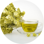
Greek Mountain Tea
Greek mountain tea, (Sideritis scardica), has been a favorite beverage in the mountainous regions of the Mediterranean for generations. It thrives in harsh growing conditions, and those challenges are what helps the plant create its protective, beneficial compounds, including polyphenols…
- The acute and chronic cognitive and cerebral blood flow effects of a Sideritis scardica (Greek mountain tea) extract: a double blind, randomized, placebo controlled, parallel groups study in healthy humans.
Background: The presence of polyphenols such as hydroxy-cinnamic acids and flavonoids in Sideritis scardica (Greek mountain tea) are likely responsible for the cognitive and mood effects of its consumption and this could be underpinned by the ability of such polyphenols to prevent monoamine neurotransmitter reuptake and to increase cerebral blood flow (CBF).
Objective: The current study extends the small amount of Sideritis scardica literature in humans by assessing both cognitive and mood outcomes in a sample of older adults, as well as blood pressure (BP) and CBF, in a subsample, utilizing near-infrared spectroscopy (NIRS).
Design: This randomized, double-blind, placebo-controlled, parallel groups trial randomized N = 155, 50–70-year-old male and female participants who were assessed for the cognitive (N = 140), mood (N = 142), BP (N = 133) and CBF (N = 57) effects of two doses of Greek mountain tea (475 and 950 mg) as well as an active control of 240 mg Ginkgo biloba, and a placebo control, following acute consumption (Day 1) and following a month-long consumption period (Day 28).
Results: Relative to the placebo control, 950 mg Greek mountain tea evinced significantly fewer false alarms on the Rapid Visual Information Processing (RVIP) task on Day 28 and significantly reduced state anxiety following 28 days consumption (relative also to the active, Ginkgo control). This higher dose of Greek mountain tea also attenuated a reduction in accuracy on the picture recognition task, on Day 1 and Day 28, relative to Ginkgo and both doses of Greek mountain tea trended towards significantly faster speed of attention on both days, relative to Ginkgo. Both doses of Greek mountain tea, relative to placebo, increased oxygenated haemoglobin (HbO) and oxygen saturation (Ox%) in the prefrontal cortex during completion of cognitively demanding tasks on Day 1. The higher dose also evinced greater levels of total (THb) and deoxygenated (Hb) haemoglobin on Day 1 but no additional effects were seen on CBF on Day 28 following either dose of Greek mountain tea. Ginkgo biloba led to lower levels of Ox% and higher levels of Hb on Day 1 and lower levels of both HbO and THb on Day 28.
Conclusions: The significantly improved cognitive performance following Greek mountain tea on Day 1 could be due to significant modulation of the CBF response. However, these improvements on Day 28 are more likely to be due to the reductions in state anxiety and, taken together, suggests that the former mechanism is more likely to facilitate acute cognitive effects and the latter more likely to underpin more prolonged cognitive improvements. [Wightman EL, Jackson PA, Khan J, et al. The acute and chronic cognitive and cerebral blood flow effects of a Sideritis scardica (Greek mountain tea) extract: a double blind, randomized, placebo controlled, parallel groups study in healthy humans. Nutrients. 2018;10:955.] - Effect of an herbal extract of Sideritis scardica and B-vitamins on cognitive performance under stress: a pilot study.
Chronic stress can impair cognitive functions including learning and memory. The current study investigated the reduction of (mental) stress and improvement of stress tolerance in 64 healthy men and women after six weeks intake of a dietary supplement containing an extract of Sideritis scardica and selected B-vitamins. Mental performance and visual attention were measured by Trail-Making Test (TMT) and Colour-Word-Test (CWT)before/after an acute stress stimulus (noise, CW-Interference). TMT improved upon product intake. The CWT reaction time accelerated upon product intake in situations of CW-Congruence (overall) (p=0.014), CW-conflict (overall) (p=0.024), CW-conflict (with noise) (p=0.001), CW-Congruence (without noise) (p=0.004) and CW-conflict (without noise) (p=0.017).CWT-changes upon product intake, differentiated for noise and CW-interference, showed (i) a bisection of CW-interference-related impairment of the reaction time in the presence of noise from 27 ms to 13.5 ms, (ii) a bisection of noise-related impairment of the reaction time in the presence of CW-conflict from 34 ms to 17 ms, (iii) an improvement of the impairment of the reaction time due to combined stress (noise plus CW-conflict) by 14.5 ms from 66 ms to 51.5 ms, (iv) despite of the improvement of the reaction time, no increase of the error rate. Safety blood parameters and the reporting of no adverse events argue for the product’s safety. These results may be relevant for persons solving cognitive tasks under conflict and/or noise (e.g. open-plan offices or car-driving) and support that the tested product alleviates stress-induced impairment of executive functioning (working memory, cognitive flexibility, controlled behavioural inhibition). [Behrendt I, Schneider I, Schuchardt JP, Bitterlich N, Hahn A. Effect of an herbal extract of Sideritis scardica and B-vitamins on cognitive performance under stress: a pilot study. Int J Phytomed. 2016;8:95-103. - Psychophysiological effects of Sideritis and Bacopa extract and three combinations thereof – a quantitative EEG study in subjects suffering from mild cognitive impairment (MCI).
Mild cognitive impairment (MCI) is regarded as a transitional stage during the development of Alzheimer’s disease. Diagnosis of MCI can be obtained by the questionnaire “DemTect” in German speaking countries. Quantitative assessment has been successfully performed using psychometric testing concomitantly with quantitative EEG recording. The present investigation aimed at the possible treatment of MCI with two botanicals, namely extracts from Sideritis scardica (500 mg) or Bacopa monnieri (320 mg) and three combinations thereof using this method in order to find a new treatment. The performance of the d2-test, an arithmetic calculation test (CPT) and a memory-test revealed better performance for the d2-test only in the presence of Sideritis extract or the combinations with Bacopa extract. Quantitative EEG assessment during the different experimental conditions showed massive differences between both extracts. Whereas Sideritis extract and its combination with a low amount of Bacopa extract (160 mg) induced increases of spectral power in fronto-temporal brain areas, Bacopa and the combination of Sideritis with high amounts of Bacopa extract produced attenuation of all waves except for delta in fronto-temporal brain areas. These differences were also documented by quantitative EEG maps in comparison to Placebo. A different action of both extracts was confirmed by discriminant analysis, where Sideritis extract and its combination with low Bacopa grouped together quite at distance to Bacopa and the combination of Sideritis with high Bacopa. A combination of Sideritis extract with a low amount of Bacopa should be tested with daily repetitive dosing for at least 4 weeks as a consequence. [Dempfel W, Schombert L, Biller A. Psychophysiological effects of Sideritis and Bacopa extract and three combinations thereof – a quantitative EEG study in subjects suffering from mild cognitive impairment (MCI). Adv Alz Dis. 2016;5:1-22. - Psychophysiological effects of a combination of Sideritis and Bacopa extract in 32 patients suffering from mild cognitive impairment. A double-blind, randomized, placebo-controlled, 2-armed study with parallel design.
Mild cognitive impairment (MCI) can be regarded as a non-demented transitional stage during the development of Alzheimer’s disease. Early recognition of this stage might increase the chance of prevention by early treatment. Within a pilot study, two plant-derived preparations and mixtures thereof were tested successfully in subjects suffering from MCI. A combination of Sideritis scardica and Bacopa monnieri extract (memoLoges®) was chosen now for a repetitive dosing during 4 weeks. Thirty-two subjects aged 50 to 80 years and suffering from MCI (having a DemTect questionnaire score between 8 and 13) were recruited for intake of 2 capsules of the preparation per day. Quantitative EEG recording during relaxation and concomitant performance of three 5 minutes lasting psychometric tests (d2-concentration test, arithmetic calculation test and memory test) was achieved at the first day and one day after the last repetitive intake. Seventeen channels of EEG and one channel EOG (for artefact rejection) were recorded. After frequency analysis (FFT) current source density was calculated as reported earlier. One, two and three hours after intake of the herbal extract or placebo the whole procedure was repeated. Brain imaging was achieved by conversion of numerical values of spectral EEG power into spectral colors and additive color mixture according to RGB as used in TV settings. Intake of memoLoges® induced a trend of improvement of performance in psychometric testing (all three tests). During relaxation quantitative as sessment of EEG data revealed attenuation of delta and theta spectral power in frontal brain as likewise reported in the presence of the Alzheimer drug rivastigmine, bringing the spectrum back to “normality”. During mental work memoLoges® induced statistically significant increases of beta power. Since MCI subjects produce less beta power in comparison to healthy subjects, this increase must likewise be seen as a positive effect pointing to a healthier spectrum. [Dimpfel W, Biller A, Suliman S, Dipah GNC. Psychophysiological effects of a combination of Sideritis and Bacopa extract in 32 patients suffering from mild cognitive impairment. A double-blind, randomized, placebo-controlled, 2-armed study with parallel design. Adv Alz Dis. 2014;5:103-125. - Assessment of the acute and subchronic toxicity and mutagenicity of Sideritis scardica Griseb. extracts.
Sideritis scardica Griseb. has a long history of collection from the wild as a traditional remedy for respiratory and gastrointestinal complaints. It has also been investigated for its promising pharmacological activities in the central nervous system. However, its toxicological data is entirely missing. This study investigated the acute and repeated-dose oral toxicity of a S. scardica 20% (v/v) ethanol extract in Sprague Dawley rats, and mutagenicity using the Ames test. No gross pathological abnormalities and no toxicity signs or mortality were detected in animals treated with the dose of 2000 mg/kg bw during 14 days of observation. The tested extract was assigned to category 5 of the GHS. To evaluate a repeated-dose toxicity, an extract has been tested over a 28-day period followed by a 14-day recovery period. No mortality and no changes in body/organ weight or food consumption have been observed. The no-observed-adverse-effect-level of the extract was determined at 1000 mg/kg bw. The results of Ames tests conducted on extracts of different polarity (water; 20% (v/v) ethanol; 50% (v/v) ethanol; n-heptane), were unequivocally negative. The study reveals no toxicity of S. scardica and no concerns for its mutagenic effects, supports its positive safety profile, and confirms the acknowledged traditional medicinal use in human. [Feistel B, Wegener T, Rzymski P, Pischel I. Assessment of the acute and subchronic toxicity and mutagenicity of Sideritis scardica Griseb. extracts. Toxins. 2018;10:258.] - Siteritis scardica extracts inhibit aggregation and toxicity and amyloid-ß in Caenorhabditis elegans used as a model for Alzheimer’s disease.
Background. Beyond its traditional uses in the Balkan area, Sideritis scardica (known as Greek mountain tea, Lamiaceae) is currently extensively investigated for its pharmacological activity in the central nervous system. Antidepressant, psychostimulating, cognition-enhancing and neuroprotective properties have been described. In this study, we tested hydroalcoholic extracts of S. scardica for their potential to counteract amyloid- _ toxicity and aggregation, which plays a crucial role in the pathogenesis of Alzheimer’s disease.
Methods. For this purpose, we have chosen the nematode Caenorhabditis elegans, which is used as a model organism for neurodegenerative diseases. The concentration of different polyphenols in extracts prepared from water, 20, 40, 50, and 70% ethanol was analysed by HPLC. Additionally, polar and unpolar fractions were prepared from the 40% ethanolic extract and phytochemically analyzed.
Results. Essentially, the contents of all measured constituents increased with the lipophilicity of the extraction solvents. Treatment of transgenic C. elegans strains expressing amyloid-_ with the extracts resulted in a reduced number of peptide aggregates in the head region of the worms and alleviated toxicity of amyloid-_, observable through the degree of paralysed animals. The mid-polar extracts (40 and 50% ethanol) turned out be the most active, decreasing the plaque number by 21% and delaying the amyloid-_-induced paralysis by up to 3.5 h. The more lipophilic extract fractions exhibited higher activity than the hydrophilic ones. Discussion. Sideritis scardica extracts demonstrated pharmacological activity against characteristics of Alzheimer’s disease also in C. elegans, supporting current efforts to assess its potential for the treatment of cognitive decline. The active principle as well as the mode of action needs to be investigated in more detail. [Heiner F, Feister B, Wink M. Siteritis scardica extracts inhibit aggregation and toxicity and amyloid-ß in Caenorhabditis elegans used as a model for Alzheimer’s disease. PeerJ. 2018;6:e4683.] - Sideritis spp. extracts enhance memory and learning in Alzheimer’s ß-amyloidosis mouse models and aged C57B1/6 mice.
Abstract. Nowadays, Alzheimer’s disease is the most prevalent epiphenomenon of the aging population. Although solubleamyloid- ß (Aß) species (monomers, oligomers) are recognized triggers of the disease, no therapeutic approach is able to stop it. Herbal medicines are used to treat different diseases in many regions of the world. On the Balkan Peninsula, at the eastern Mediterranean Sea, and adjacent regions, Sideritis species are used as traditional medicine to prevent age-related problems in elderly. To evaluate this traditional knowledge in controlled experiments, we tested extracts of two commonly used Sideritis species, Sideritis euboea and Sideritis scardica, with regard to their effects on cognition in APP-transgenic and aged, nontransgenic C57Bl/6 mice. Additionally, histomorphological and biochemical changes associated with Aß deposition and treatment were assessed. We found that daily oral treatment with Sideritis spp. extracts highly enhanced cognition in aged, non-transgenic as well as in APP-transgenic mice, an effect that was even more pronounced when extracts of both species were applied in combination. The treatment strongly reduced Aß 42 load in APP-transgenic mice, accompanied by increased phagocytic activity of microglia, and increased expression of the _-secretase ADAM10. Moreover, the treatment was able to fully rescue neuronal loss of APP-transgenic mice to normal levels as seen in non-transgenic controls. Having the traditional knowledge in mind, our results imply that treatment with Sideritis spp. extracts might be a potent, well-tolerated option for treating symptoms of cognitive impairment in elderly and with regard to Alzheimer’s disease by affecting its most prominent hallmarks: Aß pathology and cognitive decline. [Hofrichter J, Krohn M, Schumacher T, et al. Sideritis spp. Extracts enhance memory and learning in Alzheimer’s ß-amyloidosis mouse models and aged C57B1/6 mice. J Alz Dis. 2016;53:967-980.] - Greek mountain tea – an herbal drug for mental enhancement.
Herein an electroencephalographic pharmacogram (EEGP) was prepared in an animal model to detect possible psychopharmacological effects. Animals were exposed to extract of Sideritis aquos. sicc. within a crossover design vs. placebo. Single dosages were administered once a week. Strongest effects were seen with respect to alpha2 waves representing an activation of dopaminergic neurotransmission. Delta, theta, and especially at higher dosages alpha1 waves were also attenuated, compatible with the view of activation of the cholinergic, norepinephrinergic and serotonergic transmission systems. Strongest effects were always observed in the frontal cortex followed by the hippocampus. These brain areas are heavily involved in cognitive functioning. Therefore, it can be expected, that the administration of this extract also would improve mental performance. [Walbroel B, Feistel B. Greek mountain tea – an herbal drug for mental enhancement. Poster presentation. 58th International Congress and Annual Meeting of the Society for Medicinal Plant and Natural Product Research. August 29- September 2, 2010. Berlin, Germany.
Read More..
Read Less..
Hintonia Latiflora
Hintonia latiflora has been clinically studied in Germany for over 60 years in individuals with type 2 diabetes and shows impressive results. Studies have also demonstrated the ability of Hintonia latiflora to lower A1C levels by as much as 10%.
12/03/2015
-
- Hintonia concentrate: for the dietary treatment of increased blood sugar values: results of a multicentric, prospective, non-interventional study with a defined dry concentrate of hintonia latiflora.
[Translated from German] Preparations from the bark of hintonia latiflora are used to regulate the blood sugar levels. The objective of this study was to prove the nutritional benefit within the framework of a dietary treatment of increased blood sugar values with pre-diabetes and slight diabetes type 2 as well as the assessment of the tolerance and application safety. In an open, prospective, multicentric and non-interventional application study, the effects of a dry concentrate from the bark of hintonia latiflora in the form of capsules were examined for the laboratory parameters of the blood sugar levels (HbA1c, fasting and postprandial glucose) as well as for the development of diabetic accompanying symptoms (sweating, gastrointestinal symptoms, paraesthesiae, itching and neuropathies). Particular attention was also given to the tolerance and (if available) further clinical (laboratory) parameters (blood pressure, liver values and blood lipids). An eight-month treatment was documented in 178 test persons with type 2 diabetes/pre-diabetes, who were treated with oral antidiabetics and/or insulin or only with a diet. At the end of the study, 177 data records were available. The HbA1c values improved over the course of the study with a high level of clinical relevance and significance from 7.2 ± 0.4% to 6.4 ± 0.5%, in accordance with a relative improvement by 10.4% (p < 0.0001). In parallel, the values of fasting and postprandial glucose also improved by an average of 23.3 ± 12.5% (from 152.1 ± 27.4 mmol/l to 114.4 ± 18.2 mmol/l) and 24.9 ± 11.4% (from 189.5 ± 34.1 mmol/l to 140.1 ± 22.3 mmol/l). The sum score of the diabetic accompanying symptoms improved from initially 4.8 points to 1.3 points at the end of the study. Improvements were also determined in blood pressure, blood fats and liver values. The tolerance was excellent, no unwanted effects occurred, in particular no hypoglycaemic episodes. In 55 of 114 patients with antidiabetic medication (39.5%), the substance could be reduced (n = 45) or stopped entirely (n = 10). The study confirms the positive effects of the dry concentrate from the bark of hintonia latiflora on the main parameters of the blood sugar levels and the diabetic accompanying symptoms. In the event of pre-diabetes or minor cases of type 2 diabetes mellitus, this can contribute towards stabilizing the blood sugar homeostasis in particular, achieving a lower load from accompanying medication and deferring the necessity of using oral antidiabetic drugs and / or insulin. [Schmidt M, Hladikova M. Hintonia concentrate—for the dietary treatment of increased blood sugar values: Results of a multicentric, prospective, non-interventional study with a defined dry concentrate of hintonia latifloa. Naturheilpraxis mit Naturmedizin. February 2014.] - Treatment of mild and moderate type-2 diabetes: open prospective trial with Hintonia latiflora extract. Background: Extracts from the bark of Hintonia latiflora are used as di tetic measures to support the regulation of glucose metabolism and the stabilization of blood glucose values. Methods: A dry concentrated extract from the bark of Hintonia latiflora in capsule form was tested in an open, prospective clinical study in 41 dietetically stabilized subjects with type 2 diabetes. The effects on parameters of blood glucose control were documented over a period of six months. Results: Fasting and postprandial glucose and the HbA1c value declined significantly. In the case of HbA1c, this meant a reduction of the absolute value from 7.49 ± 0.72% to 6.82 ± 0.67% (from 58.4 to 51.0 mmol/mol Hb; intention to treat (ITT) population). Furthermore, cholesterol and triglycerides were slightly reduced and no negative effect on other laboratory parameters and no change of the liver values were observed. Tolerance was very good. In particular, no side effects and no hypoglycemic episodes or worsening of diabetic symptoms occurred. Conclusions: The study confirms the positive effect of extracts from the bark of Hintonia latiflora on blood glucose values suggesting a potential benefit in the management of glucose metabolism in cases of type 2 diabetes.[Korecova M, Hladikova M. Treatment of mild and moderate type-2 diabetes: open prospective trial with Hintonia latiflora extract. European Journal of Medical Research. 2014;19(1):16.]
- Hintonia latiflora in patients with type 2 diabetes: A long-term study. [Hintonia latiflora bei Typ-2-Diabetes.] The antidiabetic efficacy and safety of an extract from Hintonia latiflora (drug-extract ratio 1:4.5, extraction solvent 32% ethanol) was tested in a monocenter open, uncontrolled study on 30 patients with type 2 diabetes. Patients were adjusted to a stable diet at least two months prior to therapy and during the entire duration of the study in order to rule out a dietary impact on the study outcome. After 12 months of treatment, fasting blood glucose was reduced by 20.6%, postprandial glucose by 19% and the mean HbA1c by 10.3% (p < 0.001). In the patients remaining in the study, these parameters were stable thereafter (up to 33 months of treatment). There were no hypoglycemic episodes or adverse events throughout the entire treatment period. [Korecova M, Hladicova M, Korec R. Hintonia latiflora bei Typ-2-Diabetes. Zeitschrift für Phytotherapie. 2006;27:272-278.]
- Natural treatment of Type II diabetes utilizing an herbal antidiabetic product. Case Report. [Naturheilkundliche Therapie des Typ-II-Diabetes mit Hilfe des Phyto-Antidiabetikums.] One female patient with diabetes mellitus received an 8-week course with 5×50 Hintonia drops daily. Fasting glucose decreased in a female patient after a three-week treatment with Hintonia after a prior exacerbation of glycaemic control, despite the use of metformin as an oral antidiabetic. She was initially considered for insulin treatment. During her 8-week course with 5×50 Hintonia drops daily, the fasting glucose level was reduced from 191 mg to 176 mg. No adverse reactions were observed. [Ploss O. Naturheilkundliche Therapie des Typ-II-Diabetes mit Hilfe des Phyto-Antidiabetikums. HP Naturheilkunde. 2002;25(1/2):40-44.
- Therapeutic trial with Copalchi [Hintonia latiflora] bark in patients with impaired glucose tolerance. [Therapieversuch mit Copalchi-Rinde bei pathologischer Glucosetoleranz.] The effect of 2 – 3×30 Hintonia drops on glucose tolerance was examined on a group of fourteen subjects with impaired glucose tolerance not treated with oral antidiabetics or insulin. Although an effect of an 8-week onwards intake of the investigational medicinal product exhibited no immediate effect on the glucose tolerance test, positive long-term effects were seen in at least 7 patients (treated for several years) because none of these cases developed full diabetes. Diabetes is a progressive disease and the necessity for the adjustment to antidiabetic drugs is merely a matter of time. The delaying of the time point of an antidiabetic therapy may be considered to be an important prophylactic factor. No adverse reactions were recorded and reported [Machens R. Therapieversuch mit Copalchi-Rinde bei pathologischer Glucosetoleranz. Erfahrungsheilkunde. 1996;45(9):605-608.]
- A long-term test with Copalchi fluid extract, a herbal anti-diabetic drug. [Eim Langzeitversuch mit Copalchi-Fluidextrakt, einem pflanzlichen Antidiabetikum.] Open long-term study. Fourteen patients with type 2 diabetes received approximately 10 drops of a copalchi (Hintonia latiflora) extract twice daily for a duration of 10 months. The patients were stabilized to glibenclamide and diet prior to the administration of copalchi drops. In 9 patients “good” to “very good” improvement of their HbA1c –values was found and in 4 of these patients glibenclamide could be discontinued within 10 months. With regard to safety, no pathologic changes in liver parameters during long-term application of copalchi drops in 16 subjects with diabetes mellitus were detected. [Machens R. Eim Langzeitversuch mit Copal chi-Fluidextrakt, einem pflanzlichen Antidiabetikum. Erfahrungsheilkunde. 1992;6:416-420.]
- Two cases of spontaneous regression of diabetes mellitus Type 2 after treatment with a phytotherapeutic natural medicine. [Zwei Fälle spontaner Regression des Diabetes mellitus Type 2 nach Behandlung mit einem Phytotherapeutikum.] The effects of copalchi drops (Hintonia latiflora) were assessed in eight patients with diabetes mellitus. An initial increase of HbA1c values on short-term intake of 20 drops 3 times daily for 4 weeks was found. A substantial decrease in the fasting glucose level was observed in 4 of the 8 cases, whereas HbA1c -values were unaltered or increased in 7/8 patients. With regard to the relatively long half-life of HbA1c it was concluded that a measurement after only 4 weeks of copalchi intake is not likely a meaningful result. No adverse reactions were observed. [Machens R. Zwei Fälle spontaner Regression des Diabetes mellitus Type 2 nach Behandlung mit einem Phytotherapeutikum. Erfahrungsheilkunde. 1991;6:433-436.]
- Oral diabetes therapy with a copalchi extract. [Orale Diabetestherapie mit einem Eupharbiazeenextrakt.] In order to determine the effects of a liquid copalchi (Hintonia latiflora) extract, a dose of 50 drops three times daily was evaluated in healthy volunteers and diabetic patients. The investigation was performed by the use of a cross-over design on three consecutive days. Baseline glucose levels were measured in 10 healthy volunteers, in 15 patients with mild forms of diabetes mellitus, especially in those suffering type 2 diabetes, in cases of moderately severe diabetes mellitus (as judged by a daily insulin requirement of 30-40 IU), and in severe and insulin-resistant cases of diabetes type 2 and in diabetes type 1 (n=6). Results: In the ten healthy volunteers the administration of insulin (5 IU) distinctly lowered blood glucose levels, whereas the application of the copalchi extract (50 drops) did not produce hypoglycaemia. In the fifteen patients with mild forms of diabetes mellitus (especially in type 2 diabetes) the effects were expressed in a reduced requirement of auxiliary insulin from 15-25 IU to 5-10 IU. Insulin was discontinued in cases where the administered dose was in the range of 10-15 IU. Two reports were presented in which up to 25 IU of insulin were replaced by copalchi 50 drops/ 3 times daily with even better effects on glucose control. In cases with moderately severe diabetes mellitus (as judged by a daily insulin requirement of 30-40 IU) the insulin sparing effect was less, reaching only 5 IU insulin reduction at its maximum. A reduction in renal urine excretion was also detected. The observed effects were dose-dependent: two patients who accidentally reduced the ingested dose to 20 drops t.i.d. experienced a reoccurrence of diabetic symptoms such as thirst, diuresis and an increase of blood glucose within 1-2 days. In both cases the resetting of the dose to the initially recommended quantity of 50 drops t.i.d completely restored these effects. In severe and insulin-resistant cases of diabetes type 2 and in diabetes type 1 (n=6) the blood glucose-lowering effect was far less pronounced or missing, as demonstrated in two case reports. Co-symptoms of diabetes such as pruritus or thirst were effectively counteracted by the intake of the investigational extract. Adverse reactions were not recorded in any of the 31 cases. [Kuhr R. Orale diabetestherapie mit einem Eupharbiazeenextrakt. Landarzt. 1953;29(23):542-549.]
- Clinical validation for an herbal Hintonia product. [Klinisches Gutachten über das Hintonia latifloria Produkt.] The study was undertaken to compare the influence of Hintonia drops on blood glucose level, urinary glucose and acidosis, in mild to moderate cases of diabetes, especially in patients suffering type 2 diabetes. The dosages applied to patients with mild to moderate diabetes, (especially patients suffering type 2 diabetes) ranged between 3 x 30-50 drops/day. In order to avoid an influence of the diet, only patients were tested who were compliantly adjusted to a standard diet for a minimum of 4-5 days. In five of eight cases a distinct improvement in symptoms was observed. No adverse reactions were observed in any of the described cases. [Pellegrini A. Klinisches Gutachten über das Hintonia latiflora Produkt. Sonderdruck Fa. Sippel, Konstanz. 1951:1-7.]
- Clinical case reports: the effectiveness of the oral antidiabetic Hintonia latiflora. [Bericht über die Behandlung mit dem peroralen Antidiabetikum Hintonia latiflora.] Severe cases did not satisfactorily respond to this treatment, whereas in mild to moderate cases a significant reduction of diabetic symptoms was achieved, as expressed in a distinct reduction of urinary glucose and of blood glucose. Co-morbidity caused by the diabetes, such as pruritus, nightly sweating, furunculosis and thirst resolved quickly. Even in cases of severe diabetes (in whom no noticeable reductions of blood glucose values were observed), complaints – related to early stages of acidosis such as headache, tiredness or dizziness – were reduced. No adverse effects were observed. The author concluded that especially mild and moderate cases or diabetes and type-2 diabetes can successfully be treated with Hintonia drops. In severe cases of juvenile diabetes the treatment can at least positively influence the general health condition of the patient, even though no amelioration of blood sugar values would be expected. [Schmid P. Bericht über die Behandlung mit dem peroralen Antidiabetikum Hintonia latiflora. Sonderdruck Fa. Sippel, Konstanz. 1951: 1-4.
- Review of the oral antidiabetic, Hintonia latiflora. [Erfahrungsbericht mit dem peroralen Antidiabetikum Hintonia latiflora.] Following a run-in period of at least 6 days with a standardized diet, a 6-day treatment phase with Hintonia, 30-50 drops 3-times daily was started. Thereafter, the drops were discontinued and during this post-treatment observation period insulin was replaced as needed. “Severe” cases of DM were defined as states with an acidosis and a regular excretion of >50 g of glucose with the urine. “Moderately severe” cases were defined as having no acidosis and 20-50 g of glucose in the urine, whereas “mild” cases exhibited a daily urinary excretion below 20 g glucose. None of the patients with severe diabetes responded to the treatment with Hintonia drops. In contrast, a reduction of blood sugar and glucose excretion into the urine was achieved in 9/14 patients with moderate diabetes. The effect could be maintained over the whole treatment period and lasted up to 2-3 days after discontinuation. With the exception of diarrhea in one case during the treatment period (6 days), no adverse reactions were observed. [Vida F. Erfahrungsbericht mit dem peroralen Antidiabetikum Hintonia latiflora. Med Welt. 1951;20:1623-1624.]
- Contribution to diabetes treatment. [Beitrag zur Diabetes-Behandlung.] Diabetic patients were treated with Hintonia drops (average dose: 3×30 drops/day) within a period of 4 months. The author reported that mild to moderate cases responded best to the treatment. In patients previously (and unsuccessfully) treated with diet only diabetic symptoms reverted to normal within only 14 days of treatment with Hintonia drops. Glycaemia, glucosuria and the amount of urine within 24 hours were distinctly reduced. Accompanying diabetic-related diseases and damages such as furunculosis or pruritus were completely resolved, whereas the late occurring diabetic damages such as polyneuritis or retinopathy were not influenced. The author also noted that patients immediately felt a subjective amelioration of symptoms, as expressed in an amelioration of weakness, tiredness or nervous restlessness. No adverse effects were observed. [Ritzmann H. Beitrag zur Diabetes-Behandlung. Hippokrates. 1950;21(6):161-168.]
- Experimental Animal Studies on the Hypoglycemic Effects of a Copalchi Extract. [Tierexperimentelle Untersuchungen zur hypoglykamischen Wirksamkeit eines Copalchi-Extraktes.] The hypoglycemic effects of a Copalchi extract was tested in 60 adults – male and female – Wistar rats in the presenta randomized, placebo-controlled, parallel-comparative animal study. A control group receiving only an Altromin standard chow was compared to treatment groups which received an Altromin chow enriched with Copalchi dry substance at 0.028% m/m or at 0.28% m/m. The study period was 30 days. The distinctly significant blood glucose lowering effect of the Copalchi extract could be demonstrated in both treatment groups. No significant difference in the antidiabetic effect was seen between the two concentrations. The onset of action of the Copalchi extract was observed between treatment days 1 and 3. A 20-25% decrease in the blood glucose concentration was achieved between the 7th and 15th day of treatment and remained at this level thereafter. This plateau was reached in the female rats already between treatment day 3 and 5. The parallel observed increase in the mean body weight was not striking and generally was the same between the test animals and the control animals. [Pinto A, Capasso A, Sorrentino L. Tierexperimentelle Untersuchungen zur hypoglykamischen Wirksamkeit eines Copalchi-Extraktes. Antidiabetika. 1997;47(7):829-833.]
- Hintonia concentrate: for the dietary treatment of increased blood sugar values: results of a multicentric, prospective, non-interventional study with a defined dry concentrate of hintonia latiflora.
Read More..
Read Less..

Omega-3
Phospholipid bound Omega-3’s from salmon contain peptides to protect delicate blood vessels in the brain by fighting oxidative damage.
July 2016
Abstracts on omega-3s from salmon
-
- Anxiolytic-like effect of a salmon phospholipopeptidic complex composed of polyunsaturated fatty acids and bioactive peptides.
A phospholipopeptidic complex obtained by the enzymatic hydrolysis of salmon heads in green conditions, exert anxiolytic-like effects in a time and dose-dependent manner, with no affection of locomotor activity. This study focused on the physic-chemical properties of the lipidic and peptidic fractions from this natural product. The characterization of mineral composition, amino acid and fatty acids was carried out. Stability of nanoemulsions allowed us to realize a behavioral study conducted with four different tests on 80 mice. This work highlighted the dose dependent effects of the natural complex and its various fractions over a period of 14 days compared to a conventional anxiolytic. The intracellular redox status of neural cells was evaluated in order to determine the free radicals scavenging potential of these products in the central nervous system (CNS), after mice sacrifice. The complex peptidic fraction showed a strong scavenging property and similar results were found for the complex as well as its lipidic fraction. For the first time, the results of this study showed the anxiolytic-like and neuroprotective properties of a phospholipopeptidic complex extracted from salmon head. The applications on anxiety disorders might be relevant, depending on the doses, the fraction used and the chronicity of the supplementation. [Belhaj N, Desor F, Gleizes C, et al. Anxiolytic-like effect of a salmon phospholipopeptidic complex composed of polyunsaturated fatty acids and bioactive peptides. Marine Drug. 2013;11:4294-4317.] - Polar lipids: n-3 PUFA carriers for membranes and the brain: Nutritional interest and emerging processes.
The n-3 fatty acids are unanimously considered to have high nutritional value, especially the long chain (LC) polyunsaturated fatty acids (PUFA) from marine origin. However, most of the products available in the market contain LC-PUFA extirified on the glycerol under the common form of triglycerols. The study investigates a process by which the PUFA can be esterified on polar lipids, especially phospholipids. The patented process is performed under low temperature without use of solvents and produces a Phospho-Lipo-Peptidic Complex that is particularly rich in DHA and is esterified in the proper sn-2 position on PL. Further, it has been demonstrated that n-3 fatty acids esterified on polar lipids instead of triglycerols, and retained in the proper, natural sn-2 position, have dramatically enhanced gastrointestinal absorption and intracellular incorporation (Bourre 2004). In pharmacokinetically validated CACO-2 studies, n-3 PUFA absorption across the enteric barrier was 5 fold over triglycerol bound n-3 PUFA fish oil. Subsequent intracellular levels were 50 fold greater. Phospholipids themselves have been shown in other studies (Kidd 1999; Bernoud, et al, 1999) to have direct brain impact, especially in the area of memory and learning performances in the aging human. Therefore, a process by which n-3 PUFA can be delivered in the correct sn-2 position and esterified to phospholipids may be a preferred delivery model over n-3 PUFA delivery esterified to triglycerols in fish oil. [Parmentier M, Mahmoud C, Linder M, Fanni J. Polar lipids: n-3 PUFA carriers for membranes and the brain: Nutritional interest and emerging processes. Oléagineux, Corps Gras, Lipides; Volume 14 (Issue 3): p.224-229.]
ADDITIONAL INFORMATION
Marine Phospho-Lipo-Peptidic complex for health benefits. In recent years, marine phospholipids represent a new source of n-3 fatty acids, in particular DHA for application in foods, neutraceuticals and cosmetics. According to the commercial terminology marine lecithin is used as a broad definition to describe a mixture of polar lipids (phospholipids and glycolipids) comprising predominantly long chain and highly unsaturated fatty acids obtained from a marine raw material. Phospholipids are major constituents of biological cell membranes. These amphiphilic molecules have a structural and functional synergy with fatty acids. Those are the lipophilic moiety associated with an hydrophilic part in form of a phosphoric acid ester. Phospholipids mainly differ by the group linked to the phosphoric ester (e.g. choline, ethanolamine, inositol, serine, …). The metabolic efficiency of these molecules depends on the type and location of the unsaturation of fatty acids. Marine phospholipids are characterized by very high levels of mostly long chain and highly unsaturated fatty acids mainly eicosapentaenoic and docosahexaenoic acids (C20:5 n-3 and C22:6 n-6, respectively). Docosahexaenoic acid (DHA), the end product of the omega-3 family fatty acid, is an abundant component in the brain phospholipids and a major nutrient of marine lipids. DHA is particularly enriched in the brain, retina, and spermatozoa phospholipids and plays a crucial role in brain development, learning ability, and visual acuity. [Linder M. Marine phosphor-lipo-peptidic complex for health benefits. White paper.]Analysis of lipids extracted from salmon (Salmo salar) heads by commercial proteolytic enzymes. Fresh salmon heads were submitted to controlled proteolysis using food-grade commercial enzymes (Alcalase, Neutrase, and Protamex). The release of oil under mild conditions (60°, 2h) compared favourably with organic solvent extraction (1.98% vs. 21.5%). Lipids extracted by solvent and lipids resulting from enzymatic processes displayed a similar content of PUFA (about 35%), mainly eicosapentaenoic acid (EPA; 8.4% vs. 7.7%) and docosahexaenoic acid (DHA; 12.1% vs 11.9%). Thin-layer chromatography (TLC-FID latroscan) showed that the polar lipid fraction accounted for 55% of total lipids (phophatidylethanolamine, 20.7%; phosphatidylcholine, 14.8%). Salmon head phospholipids may be more effective carriers of highly unsaturated fatty acids to specific tissues than triacyglycerols, as shown by their content in EPA (10.3 and 6.9%, respectively) and DHA (33.1 and 9.1%, respectively). [Gbogouri GA, Linder M, Fanni J, Parmentier M. Analysis of lipids extracted from salmon (Salmo salar) heads by commercial proteolytic enzymes. Eur J Lipid Sci Technol. 2006;108:766-775.]
Doxosahexaenoic acid prevents neuronal apoptosis induced by soluble amyloid-β oligomers. A growing body of evidence supports the notion that soluble oligomers of amyloid-β (Aβ) peptide interact with the neuronal plasma membrane, leading to cell injury and inducing death-signaling pathways that could account for the increased neurodegeneration occurring in Alzheimer’s disease (AD). Doxosahexaenoic acid (DHA, C22:6, n-3) is an essential polyunsaturated fatty acid in the CNS and has been shown in several epidemiological and in vivo studies to have protective effects against AD and cognitive alterations. However, the molecular mechanisms involved remain unknown. We hypothesized that DHA enrichment of plasma membranes could protect neurons from apoptosis induced by soluble Aβ oligomers. DHA pre-treatment was observed to significantly increase neuronal survival upon Aβ treatment by preventing cytoskeleton perturbations, caspase activation and apoptosis, as well as by promoting extracellular signal-related kinase (ERK)-related survival pathways. These data suggest that DHA enrichment probably induced changes in neuronal membrane properties with functional outcomes, thereby increasing protection from soluble Aβ oligomers. Such neuroprotective effects could be of major interest in the prevention of AD and other neurodengenerative diseases. [ Florent S, Malaplate-Armond C, Youssef I, et al. Doxosahexaenoic acid prevents neuronal apoptosis induced by soluble amyloid-β oligomers. J Neurochem. 2006;96:385-395.]
- Anxiolytic-like effect of a salmon phospholipopeptidic complex composed of polyunsaturated fatty acids and bioactive peptides.
Read More..
Read Less..
Pleuran from Oyster Mushroom
1. Beneficial effects of pleuran on asthma control and respiratory tract-infection frequency in children with perennial asthma.
The aim of this study was to evaluate the effects of pleuran (β-glucan isolated from Pleurotus ostreatus) on asthma control and respiratory morbidity in children on conventional GINA-based asthma treatment who had partially controlled perennial asthma. A double-blind, placebo-controlled multicentre clinical trial with a 2-arm, parallel design was conducted across three countries; 230 children aged 7 to 17 years were randomised (1:1) into an active group (receiving a pleuran/vitamin C combination) or a placebo group (receiving vitamin C only). This study consisted of 24 weeks of treatment (2 capsules a day) and then 24 weeks of follow-up. The primary endpoints included the effects of active treatment versus placebo on asthma control and respiratory tract infections (RTIs). Secondary endpoints included changes in the following measures: number of asthma exacerbations, with or without respiratory infection; quality of life of both asthmatic children and their caregivers; spirometric indices; fractional exhaled nitric oxide (FeNO) levels; safety after 24 weeks of treatment and also after the full 48-week study period. Overall, 206 children completed this study; 113 of these children were in the active group and received a pleuran/vitamin C combination for 24 weeks. After the 24-week treatment period, children below 12 years of age who were in the active group achieved significant improvements in asthma control compared to those in the placebo group (21.8 ± 3.5 vs. 20.3 ± 4.0; P = 0.02); while children at least 12 years old who were in the active group reported lower numbers of RTIs (0.7 ± 1.0 vs. 1.9 ± 1.7; P = 0.002) compared to children of this age in the placebo group. In addition, children below 12 years of age in the active group showed a significant decrease in asthma exacerbations compared to those in the placebo group (2.5 ± 1.6 vs. 3.3 ± 1.9; P = 0.05). At the end of the 48-week trial, a statistically significant improvement in asthma control was observed in 84.7% of children who received pleuran/vitamin C treatment compared to 67.0% of children who received vitamin C only (P = 0.01). The pleuran/vitamin C combined treatment was safe and well-tolerated, and no related serious adverse events were reported. This study highlights the favourable safety profile of pleuran/vitamin C supplementation and demonstrates positive effects of this treatment on asthma control and RTI incidence in children with allergic perennial asthma that was partially controlled by conventional therapy. [Jesenak M, Hrubisko M, Chudek J, et al. Sci Rep. 2025;15:7146.]
2. Effect of a pleuran-based supplement on salivary IgA secretion in children with recurrent respiratory infections.
Background: ß-glucans isolated from natural sources have demonstrated pluripotent immunomodulatory potential, making them a promising supportive treatment for the management of recurrent respiratory infections (RRIs) in children. This study aimed to evaluate the effects of a pleuran-based supplement (ß-glucan isolated from Pleurotus ostreatus in combination with vitamin D and zinc) on mucosal immunity -through modulating salivary secretory immunoglobulin A (sIgA) levels -in children with RRIs. Methods: This monocentric, prospective, open-label pilot study investigated the effect of an orally administered pleuran/vitamin D/zinc supplement (1-2 chewable tablets daily depending on body weight) on the dynamics of sIgA secretion measured in saliva samples collected at three timepoints: at baseline and after 4-6 and 8-10 days. Results: This study included 49 children aged 6-11 years (mean age: 8.2 ± 1.6 years) with a history of one or more of the following conditions in the inclusion criteria: RRIs, allergy, and asthma. After 8-10 days with daily administration of the chewable pleuran/vitamin D/zinc supplement, children exhibited a statistically significant increase in salivary sIgA concentrations compared with baseline (227 ± 211 μg/mL; P = 0.045). No adverse events were observed during the course of the study in relation to the administration of pleuran-based supplement. Conclusions: We demonstrated the beneficial effects of the short-term administration of a pleuran-based chewable supplement on mucosal immunity through increasing salivatory sIgA levels. This study confirms the favourable safety profile of this pleuran/vitamin D/zinc combination, which could be beneficial for children with acute or recurrent respiratory infections, including children with allergies and/or asthma. Moreover, the significant increases in salivary sIgA concentrations that were observed after a few days of supplementation support the use of pleuran in not only the prevention but also the treatment of acute respiratory infections. [Kunc P, Fabry J, Matiscakova M, et al. Curr Ther Res Clin Exp. 2025;102:100780.]
3. Immunomodulation with β-glucan from Pleurotus ostreatus in patients with endocrine-dependent breast cancer.
Aim: To evaluate the effect of pleuran (β-glucan from Pleurotus ostreatus) administration on the immune profile of patients with endocrine-dependent breast cancer (clinical stages I-II) in clinical and imaging remission. Methodology: Antitumor cellular immunity (CD19+, CD3+, CD4+ and CD8+ T lymphocytes and natural killer cells) of 195 patients (49 in the pleuran group and 146 in the control group) was measured by flow cytometry. Results: We observed a significant increase in the absolute number of CD3+, CD19+, CD4+ and CD8+ T lymphocytes in the pleuran group compared with the control group. Conclusion: Our results suggest potential benefit of continuous pleuran administration on immune rehabilitation of cellular antitumor immunity and better prognosis in breast cancer patients in remission. [Spacek J, Vocka M, Zavadova E, Konopasek B, Petruzelka L. Immunotherapy. 2022;14(1):31-40.]
4. Efficacy of pleuran (β-Glucan from Pleurotus ostreatus) in the management of herpes simplex virus type 1 infection.
One of the highly prevalent viral pathogens among children and adults causing infection, clinically presenting as herpes labialis, is herpes simplex virus type 1 (HSV-1). The long-term administration of acyclovir, a standard regimen for therapy against HSV-1 infections, can cause viral resistance against this drug. Therefore, the development of natural drugs with low toxicity that are able to enhance host antiviral defense against HSV infection is needed. β-Glucans represent a type of biologically active molecules possessing antiviral properties. The goal of this study was to investigate the clinical and immunomodulatory effect of β-glucan pleuran (insoluble β-1,3/1,6-D-glucan isolated from Pleurotus ostreatus) based supplements on the duration and intensity of herpes symptoms and on the incidence rate and duration of acute respiratory symptoms and intercurrent diseases in HSV-1 positive patients. Ninety patients were randomised into active and placebo groups. Active treatment with pleuran in systemic application caused a significantly shorter duration of herpes simplex symptoms compared to the placebo group. During the preventive phase (120 days), the duration and severity of respiratory symptoms were lower in the active group compared to the placebo group; however, a significant difference was found only in the case of cough. No significant side effects were observed during both phases of the clinical trial (acute and preventive). Obtained results suggest that the use of pleuran seems to be a promising approach in the treatment of acute HSV-1 with beneficial effect on the respiratory tract symptoms and infections. [Urbancikova I, Hudackova D, Majtan Juraj, et al. Evid Based Complement Alternat Med. 2020 Apr 3:2020:8562309. doi:10.1155/2020/8562309.]
5. Nové možnosti liečby akútnych respiračných ochorení viaczložkovým prírodným prípravkom s obsahom pleuranu (biologicky aktívny polysacharid z Pleurotus ostreatus) u detí – otvorené klinické pozorovanie.
No abstract available. [Jesenak M., Urbancikova I., Hudackova D., Rennerova Z., Kuniakova R., et al. Revue medicíny v praxi. 2020, roč. 18, č. 2, s. 31-36.]
6. Effects of pleuran (β-glucan from Pleurotus ostreatus) supplementation on incidence and duration of bronchiectasis exacerbations.
BACKGROUND: Patients with non-cystic fibrosis bronchiectasis (BE) have frequent exacerbations that are causes of significant morbidity and sometimes mortality, and which it is desirable to prevent. AIM: We aimed to assess the effects of pleuran on the incidence and duration of exacerbations in patients with BE. METHODS: A prospective, observational, open-label, and active-controlled study was realized as a comparison of the frequency and duration of exacerbations between a group of patients with BE (30 patients, 14 males and 16 females, aged 44–72 years) who received a combination supplement containing pleuran 100 mg, Vitamin C 60 mg and zinc 5 mg over a 3-month period and a group of patients with BE (31 patients, 15 males and 16 females, aged 45–74 years) treated over a 3-month period with a combination supplement containing Vitamin C 60 mg and zinc 5 mg. RESULTS: Over the study period, altogether 46 exacerbations were documented (19 in the patients receiving pleuran and 27 in the patients who did not receive pleuran), nine of which required hospital treatment (four in the patients receiving pleuran [21.5%] and five in the patients who did not receive pleuran [18.6%]). The mean number of exacerbations over the study period was significantly lower in the patients receiving pleuran (0.6 ± 0.4) as compared to the mean number in the patients who did not receive pleuran (0.8 ± 0.3) (p = 0.0297). The mean duration of exacerbations, expressed in days, needed for cure or clinical improvement in the patients receiving pleuran (11.2 ± 1.7 days) was significantly shorter than that of exacerbations in the patients who did not receive pleuran (12.4 ± 1.3 days) (p = 0.0029). We found significantly lower incidence and significantly shorter duration of exacerbations in the patients with BE who received pleuran as compared to their incidence and duration in the patients with BE who did not receive pleuran. CONCLUSION: Our findings indicated a need for further investigations in this domain to define the possible role of pleuran in the prevention of BE exacerbations. [Minov J, Stoleski S, Karadzinska-Bislimovska J, et al. Open Access Maced J Med Sci. 2020;8 No. B:906-12.]
7. Effects of pleuran (Β-Glucan from Pleurotus Ostreatus) supplementation on incidence and duration of COPD exacerbations.
Background: 1,3/1,6-β-glucans are recognised as immunomodulators in human and veterinary medicine for over 50 years. Aim: To assess the effects of pleuran (1,3/1,6-β-glucan from Pleurotus ostreatus) on incidence and duration of bacterial exacerbations in patients with COPD. Methods: We performed an observational, non-randomized, open-label study including 32 COPD patients (Group D) in whom besides the recommended chronic treatment for the stable disease were administered supplement combination containing pleuran 100 mg, vitamin C 60 mg and zinc 5 mg once daily over a three month-period (Group 1). Also, an equal number of Group D COPD patients who besides the recommended treatment for stable disease received the supplement combination containing vitamin C 60 mg and zinc 5 mg once daily, matched to the study subjects of the Group 1 by sex and age served as control (Group 2). Results: Over the study period 57 exacerbations (24 in the Group 1 and 33 in the Group 2) were documented. A mean number of exacerbations over the study period was significantly lower in the Group1 (0.7 ± 0.4) as compared to their mean number in the Group 2 (1.0 ± 0.6) (P = 0.0218). Furthermore, a mean duration of exacerbations expressed in days needed for cure or clinical improvement (i.e. complete resolution of symptoms or return of the symptoms to their baseline severity) in the Group 1 (6.7 ± 0.8 days) was significantly shorter than the mean duration of exacerbations in the Group 2 (7.4 ± 1.3 days) (P = 0.0118). There was not reported any adverse effect during the study period by study subjects from both examined groups. Conclusion: Our findings indicated that pleuran might impact the incidence and duration of bacterial exacerbations in patients with COPD. There is a need for further studies for more precise determination of the influence of pleuran on the course of COPD. [Minov J, Bislimovska-Karadzhinska J, Petrova T, et al. Open Access Maced J Med Sci. 2017;5(7):893-898.]
8. Preventive effect of pleuran (β-glucan from Pleurotus ostreatus) in children with recurrent respiratory tract infections – Open-label prospective study.
Background: Recurrent respiratory tract infections (RRTIs) are one of the most common problems in pediatric practice. Several natural preparations are used in management of RRTIs, but the scientific evidence for their efficacy is sometimes insufficient. On the other hand, preventive effect of some natural preparation (e.g. β-glucans) was confirmed by relevant clinical studies. Methods: In our prospective open label study we enrolled 194 children suffering from RRTIs. We aimed to analyse the effect of Imunoglukan P4H® syrup containing pleuran (insoluble β-glucan isolated from Pleurotus ostreatus) on general respiratory morbidity comparing previous year with the same season during the study. Results: Supplementation of Imunoglukan P4H® syrup significantly decreased the total number of respiratory tract infections during the treatment and subsequent follow-up period compared to the same period of the previous year (4.18 ± 2.132 vs. 8.71 ± 1.89; p<0.001). In detail, the number of various types of respiratory tract infections (otitis, laryngitis, bronchitis and common cold) was also significantly reduced (p<0.01 for all subtypes of infections). Moreover, a reduction in the number of day-off in kindergarten and school was also noticed. The syrup was well tolerated and no serious adverse effects were observed. Conclusion: Our study supports the use of pleuran in the complementary treatment and preventions of RRTIs in children. β-glucans seem to be an effective and safe tool
in the management of RRTIs. [Pasnik J, Slemp A, Cywinka-Bernas A, et al. Curr Ped Res. 2017;21(1):99-104.]
9. Beta-(1,3/1,6)-D-glucan helps to decrease opportunistic infections in Crohn’s disease patients treated with biological therapy.
Background: Secondary intercurrent infections in patients with inflammatory bowel disease (IBD) represent a very important problem in daily clinical practice because of their significant contribution to quality of life and work ability. Beta-glucans are known to act as non-specific immunomodulators that exhibit an anti-inflammatory effect as evident in animal and human studies. Imunoglukan P4H® capsules are nutritional supplement containing 100 mg of beta-(1,3/1,6)-D-glucan pleuran isolated from oyster mushroom Pleurotus ostreatus and 100 mg of vitamin C. Aims: To evaluate the influence of Imunoglukan P4H® (Pleuran, s.r.o., Slovakia) on the secondary morbidity added to biological therapy in maintaining remission of Crohn´s disease (CD) for 12 months. Patients and methods: A double-blinded, placebo-controlled, randomised bicentric study was carried out which included 53 CD patients randomly divided into two groups treated with Imunoglukan P4H® (with beta-(1,3/1,6)-D-glucan pleuran from Pleurotus ostreatus and vitamin C) 1 capsule daily (23 patients) and placebo (vitamin C only) 1 capsule daily (30 patients) respectively. All patients also continued their biological maintenance therapy. Before inclusion the patients were required to: be consented (written), complete a standard quality of life questonnaire for patients with inflammatorybowel disease (SIBDQ), Crohn´s disease activity index (CDAI) was evaluated, full blood count, standard biochemical tests – bilirubine (Bi), alaninaminotranspherase (ALT), aspartataminotranspherase (AST), alcaline phosphatase (ALP), gammaglutamyltranspeptidase (GMT), serum amylases (AMS), natrium (Na), kalium (K), C-reactive proteine (CRP) and faecal calprotectin were also performed. These tests and questionaires were repeated at 2-month intervals, at each time patients reported intercurrent diseases and their lengths. All data were recorded in patient protocol and statistical signification was tested by matching of two ratios with Yates correction and by Mann-Whitney test. Results: From 53 included CD patients 39 reached the end of trial, 14 patients interrupted the study prematurely. Imunoglukan P4H® was administered to 10 patients and placebo to 29 patients. 0/10 patients in Imunoglukan P4H® group (IG) and 1/29 patients in placebo group (PG) interrupted rematurely administration of preparation for intolerance and 1/10 patients in IG and 2/29 patients in PG for relapsing of CD. These differences were not statistically significant (p=0,5519 and 0,7508 resp.). At least one intercurrent infectious or allergic disease occurred in 6/10 of IG patients and in 25/29 of PG patients. In 4/10 respectively 11/29 of patients one intercurrent disease, in 2/10 resp. 7/29 of patients two, in 0/10 resp. 5/29 of patients three and in 0/10 resp. 2/29 of patients four diseases were reported. Imunoglukan P4H® significantly decreased the number of intercurrent diseases compared to placebo in this study (95 % CI, p=0,0196). The treatment did not influence SIBDQ, CDAI, blood count and biochemical parameters tested. Conclusion: Imunoglukan P4H® added to biological therapy in CD patients in remission has significantly reduced the secondary morbidity in comparison to placebo. This preparate was well tolerated, no adverse or undesirable effects were observed and the course of CD remission remained unchanged. [Batovsky M, Tomas Z, Khaled R, et al. Arch Clin Gastroenter. 2015:5-8.]
10. Anti-allergic effect of pleuran (β-glucan from Pleurotus ostreatus) in children with recurrent respiratory tract infections.
Recurrent respiratory tract infections (RRTIs) present a very important problem in paediatric praxis. As true immunodeficiencies are rare, one of the most important factors assumed to contribute to increased respiratory morbidity is atopy. Several preparations of natural origin have been used for the prevention of RRTIs, and some of the most effective immunomodulators are biologically active polysaccharides – e.g. ß-glucans. In our randomised, double-blind, placebo-controlled study, we investigated the prevalence of atopy in a group of children with RRTIs and the potential anti-allergic effect of pleuran (ß-glucan isolated from Pleurotus ostreatus) on basic laboratory markers of allergic inflammation. We confirmed that atopy may be an important factor contributing to the increased respiratory morbidity in children with RRTIs. The active treatment with pleuran resulted in a significant reduction of peripheral blood eosinophilia and stabilised the levels of total IgE in serum. This was more evident in atopic subjects. Pleuran showed a potential anti-allergic effect. This previously non-described effect could expand the application of this natural immunomodulator also as a complementary adjuvant therapy in allergic patients. [Jesenak M, Hrubisko M, Majtan J, Rennerova Z, Banovcin P. Phytother Res. 2014;28(3):471-4.]
11. Pleuran (β-glucan from Pleurotus ostreatus) supplementation, cellular immune response and respiratory tract infections in athletes.
Prolonged and exhausting physical activity causes numerous changes in immunity and sometimes transient increases the risk of upper respiratory tract infections (URTIs). Nutritional supplements as countermeasures to exercise-induced changes have increasingly been studied in the last decade. One of the most promising nutritional supplements is β-glucan, a well-known immunomodulator with positive effects on the function of immunocompetent cells. In this double blind, placebo-controlled study, we investigated the effect of pleuran, an insoluble β-(1,3/1,6) glucan from mushroom Pleurotus ostreatus, on selected cellular immune responses and incidence of URTI symptoms in athletes. Fifty athletes were randomized to pleuran or placebo group, taking pleuran (commercial name Imunoglukan(®)) or placebo supplements during 3 months. Venous whole blood was collected before and after 3 months of supplementation and additionally 3 months after supplementation period was completed. Incidence of URTI symptoms together with characterization of changes in phagocytosis and natural killer (NK) cell count was monitored during the study. We found that pleuran significantly reduced the incidence of URTI symptoms and increased the number of circulating NK cells. In addition, the phagocytosis process remained stable in pleuran group during the study in contrast to placebo group where significant reduction of phagocytosis was observed. These findings indicate that pleuran may serve as an effective nutritional supplement for athletes under heavy physical training. Additional research is needed to determine the mechanisms of pleuran function. [Bergendiova K, Tibenska E, Majtan J. Eur J Appl Physiol. 2011;111(9):2033-40.]
12. Effect of Pleuran (β-glucan from Pleurotus ostreatus) supplementation on cellular immune response after intensive exercise in elite athletes.
Excessive and exhausting physical loads depress the immune system. Carbohydrate consumption may minimize the postexercise suppression of the innate immune system. β-Glucan is a well-known immunomodulator, with positive effects on the functioning of immunocompetent cells. The goal of this study was to determine whether β-glucan dietary supplementation from the mushroom Pleurotus ostreatus decreases the suppressed immune system responses induced by short-term high-intensity exercise in humans. In this double-blind pilot study, 20 elite athletes were randomized to β-glucan (n = 9) or placebo (n = 11) groups; these groups consumed 100 mg of β-glucan (Imunoglukan) or placebo supplements, respectively, once a day for 2 months. Venous whole blood was collected before and after 2 months of supplementation (baseline), both immediately and 1 h after (recovery period) a 20-min intensive exercise bout at the end of the supplementation period. The blood samples were used to measure the cell counts of leukocytes, erythrocyte, and lymphocytes; subpopulations of lymphocytes, granulocytes, and monocytes; and natural killer (NK) cell activity (NKCA). A 28% reduction in NKCA (p < 0.01) below the baseline value was observed in the placebo group during the recovery period, whereas no significant reduction in NKCA was found in the β-glucan group. In addition, no significant decrease in NK cell count was measured in the β-glucan group during the recovery period. Immune cell counts did not differ significantly between the groups. These results indicate that insoluble β-glucan supplementation from P. ostreatus may play a role in modulating exercise-induced changes in NKCA in intensively training athletes. [Bobovcak M, Kuniakova R, Gabriz J, Majtan J. Appl Physiol Nutr Metab. 2010 Dec;35(6):755-62.]
13. Immunomodulatory effect of pleuran (β-glucan from Pleurotus ostreatus) in children with recurrent respiratory tract infections.
Objectives: Recurrent respiratory tract infections (RRTIs) represent a very important problem in daily clinical practice because of their significant contribution to morbidity in children. Several natural nutritional supplements have been used in the prevention of RRTIs, but the clinical efficacy of only a few preparations is supported by scientific evidence. Materials and methods: In a double-blind, placebo-controlled, randomised, multicentre study, we have observed a group of 175 children (aged 5.65 ± 2.39 years) with more than 5 respiratory infections that occurred during the 12 months prior to the beginning of the study. Children were randomised into an active group, treated with Imunoglukan P4H® syrup (with pleuran-β-glucan from Pleurotus ostreatus and vitamin C), or a placebo group (vitamin C only). During the 3 visits, within a 12-month period, questionnaires were completed, and blood samples were examined for immune parameters. Results: In the active group, 36% of the children did not suffer from any respiratory infections throughout the treatment, compared to 21% in the placebo group (p<0.05). Imunoglukan P4H® also significantly decreased the frequency of flu and flu-like disease and the number of lower respiratory tract infections. Imunoglukan P4H® treatment resulted in a statistically significant modulation of humoral and cellular immunity. Conclusions: Results from this study demonstrate that Imunoglukan P4H® is effective in the prevention of RRTIs in children. Furthermore, our results also revealed complex immunomodulatory activity of this product. This is the first double-blind, placebo-controlled study in children with RRTIs that has addressed the preventive effects of pleuran on morbidity caused by respiratory infections. [Jesenak M, Majtan J, Rennerova Z, Kyselovic J, et al. Int Immunopharmacol. 2013 Feb;15(2):395-9. doi: 10.1016/j.intimp.2012.11.020.]
14. Beta-(1,3/1,6)-D-glucan from Pleurotus ostreatus in the prevention of recurrent respiratory tract infections: An international, multicentre, open-label, prospective study.
Preschool children are particularly susceptible to recurrent upper and lower respiratory tract infections due to their immune immaturity and other contributing factors. Preventing and/or treating children suffering from recurrent respiratory tract infections (RRTIs) is challenging, and it is important to provide more clinical evidence about the safety and efficacy of natural immunomodulating preparations, including β-glucans. The aim of the present study was to assess the incidence of respiratory tract infections (RTIs) in children with a history of RRTIs for a period of 6 months (3 months of pleuran supplementation and 3 months of follow-up) compared with the same period from October to March of the previous year prior to enrolment in the study. A total of 1,030 children with a mean age of 3.49 ± 1.91 years from seven countries were included in this study. The total number of RTIs observed during the study period was significantly lower compared to the same period of the previous year (7.07 ± 2.89 vs. 3.87 ± 3.19; p < 0.001). Analysis of each type of RTI revealed significant reductions in the mean number and duration of infections for all RTI subtypes compared to the previous year. This study also confirmed the beneficial safety profile of pleuran supplementation. In conclusion, pleuran supplementation represents an interesting and prospective supplement in preventing respiratory infections and reveals new strategies for supporting immune functions in the paediatric population. [Rennerova Z, Sirvent LP, Roca EC, et al. Beta-(1,3/1,6)-D-glucan from Pleurotus ostreatus in the prevention of recurrent respiratory tract infections: An international, multicentre, open-label, prospective study. Front Pediatr. 2022;14:10:999701.]
15. Beta-glucans from Pleurotus ostreatus for prevention of recurrent respiratory tract infections.
Introduction: Respiratory tract infections (RTI) are one of the most common reasons for medical visits in paediatric clinical practice. In cases of recurrence, supplementation with natural derivatives such as β-glucans has been proposed as an immunomodulatory intervention which based on its effects on humoral and cell-mediated immune responses, might reduce frequency of respiratory tract infections (RTIs). Patients and methods: A prospective observational multicenter study of patients from 20 paediatric clinics of Alicante, Barcelona and Valencia who were considered with RRI according to the number of RTI during the previous year. Supplementation with a daily dose of a β-glucans from Pleurotus ostreatus and vitamin C for 3 months was recommended. Comparison between the number and type of RTI during the previous October-period, among other variables, were compared with those observed during the 6-month follow-up. Results: Analysis of data from 151 children have shown significant reductions on the average number of RTIs episodes (4.27 ± 2.21 vs. 8.88 ± 3.35; p <0.001) and the risk of been considered as RRI (RR [95%CI]: 0.36 [0.3-0.45]; p <0.0001) after the supplementation. Visits to the Emergency department, use of pharmacological treatment and missed school days were also less frequent when compared with the previous year. Each of the RTI subtypes (otitis, common cold, pharyngitis, laryngitis and bronchitis) showed significant reductions both on incidence and number of episodes. A 90.7% of cases reported a good or very good tolerability and the improvement perceived was reported in 85.7% of parents. Conclusions: In absence of measures for active immunization against pathogens most frequently associated with respiratory infections, prophylactic supplementation β-glucans from Pleurotus ostreatus and vitamin C has may be of benefit particularly in children with recurrences and specific risk factors. [Grau JS, Sidrvent LP, Ingles MM, Urgell MR. Acta Pediatrica Espanola. 2015;73(8):186-93.]
16. Imunoglukan P4H® v prevencii recidivujúcich infekcií dýchacích ciest v detskom veku.
Abstract not available. [Jesenak M, Kol A. Cesk Pediatr. 2010;65(11):639-647.]
17. Induction of metalloproteinase 9 secretion from human keratinocytes by pleuran (beta-glucan from Pleurotus ostreatus).
Glucan preparations, primarily modified water-soluble glucans, are involved in the activation of the body’s natural defense mechanisms and in the acceleration of the skin’s wound-healing processes. Pleuran, an insoluble beta-D-glucan in hydrogel form, offers a natural alternative to more common chemically derivated soluble beta-D-glucans. Pleuran was applied to human keratinocyte primary cultures, and after 24 h of incubation the release of matrix metalloproteinase 9 (MMP-9) and metalloproteinase 2 (MMP-2) by stimulated keratinocytes was detected using gelatine zymography. There was a concentration-dependent increase in pro-MMP-9 release after treatment with pleuran over the concentration range of 2 to 200 microg/ml, but pro-MMP-2 was detected at a constant level. Moreover, the active forms of both MMPs were not detectable, indicating that in vitro autoactivation of these zymogens did not occur. The results indicate that pleuran is a potent keratinocyte stimulator of proMMP-9 release, which implies its application in dermatological therapies. [Majtan J, Kumar P, Koller J, Dragunova J, Gabriz J. Z Naturforsch C J Biosch. 2009;64;(7-8)597-600.] Preclinical Research
18. The effect of an insoluble fungal glucan on indicators of immunity in calves
Immunomodulative effect of insoluble fungous glucan (IFG) was observed on nine accidentally chosen calves, which were in the age from 4.5-5.5 months. The following items were observed: T, B lymphocytes, white blood cell count, index and activity of phagocytosis, induction and determination of interferon from leukocytes. In the experimental group there was a decrease in B-rosseting cells observed. On the 28-th day and on the 42-th day of experiment duration a significant increase in B-rosseting cells in comparison with control group has been observed. Average values were significantly different on the levels P < 0.01 and P < 0.05. Percentages of T-rosseting cells were almost the same during the whole experiment. In the experimental group on the 7-th and on the 14-th day the decrease has been observed. By the Student’s t-test a significant difference in favour of group, where IFG was applicated, has been observed. Percentage of white blood cell count was from 47 to 72 in both groups. When phagocytic activity was evaluated, there was the higher percentage of phagocytic cells in the 7th, 14-th, 21-st, 28-th and 42-th day of experiment when compared with the 0-day value. Concerning the phagocyte index a statistical difference was observed, when compared to the initial value in favour of the experimental group. The initial disbalanced values of interferon, which have been observed during the period from the 7-th to the 21-st day, increased on the 28-th and the 42-nd day of experiment which was statistically confirmed on the following levels: P < 0.05 and P < 0.01. [Levkutova M, Bajova V, Hipikova V, et al. Vet Med (Praha). 1993;38(1):31-42.]
19. Delayed hypersensitivity and the primary antibody reaction after administration of infectious bovine rhinotracheitis inactivated oil vaccine to calves premedicated with glucan
The level of delayed skin hypersensitivity (DSH) to DNFB and of the primary immune reaction was evaluated in the calves immunized with an inactivated oil IBR vaccine (V group) and in the calves premedicated with glucan (seven days before vaccine administration) and subsequently immunized with the mentioned vaccine (GV group). The DSH test did not reveal an alteration of cellular immunological reactivity in the calves immunized with an inactivated oil IBR vaccine; after the vaccine administration there was no significant difference in the DSH level from the value before vaccine administration nor in comparison with the value of control calves (K group), Tab. I. But in the immunized calves which were glucan-premedicated (from Pleurotus ostreatus; 10 mg/kg l.w.) a significantly higher DSH level was determined not only in comparison with the initial value (P < 0.05) before glucan administration but also with the value of control calves (P < 0.05), Tab. I. The categorization of calves according to the DSH level also points to the immunomodulating effect of glucan in this sense (Fig. 1); while the value of skin test ranged from 3.6 to 6.5 mm in the highest percentage of the calves of V and K groups on the dates before and after administration of the mentioned preparations, a marked increase (on the date after administration) in the number of calves (from 22 to 67%) with the value of skin test higher than 6.5 mm was observed in the calves of GV group. The vaccine administration in itself (V group) did not induce the production of measurable amounts of serum IBR antibodies till day 14 after immunization. [Paulik S, Bajova V, Benko G. Vet Med (Praha). 1993;38(7):395-402.]
20. The immunomodulatory effect of the soluble fungal glucan (Pleurotus ostreatus) on delayed hypersensitivity and phagocytic ability of blood leucocytes in mice.
The effect of fungal and yeast glucan on different immune functions in mice was examined and compared. The simultaneous administration of glucan and a sensitizing dose of DNFB on the different sites significantly stimulated delayed-type hypersensitivity (DTH) response only when using fungal glucan. Both glucans tested, when administered before sensitization, significantly increased DTH response, but with a significantly higher level at the beginning of the investigation (on day 7) when using fungal glucan. The increase in phagocytic activity by the blood leucocytes started in the 1st week after fungal-glucan treatment, and in the 2nd week after yeast-glucan treatment, and took longer after administration of fungal glucan. The values of the phagocytic-activity index were significantly influenced only after fungal-glucan injection. The results of the study indicate that fungal glucan isolated from Pleurotus ostreatus could be a prospective immunomodulating substance. [Paulík S., Mojzisova J., Durove A., Benisek Z., Huska M. Zentralbl Veterinarmed B. 1996 May;43(3):129-35.]
21. Effect of pleuran (beta-glucan from Pleurotus ostreatus) on the antioxidant status of the organism and on dimethylhydrazine-induced precancerous lesions in rat colon.
The effect of pleuran (beta-1,3-D-glucan isolated from the oyster mushroom Pleurotus ostreatus) on the antioxidant status of the organism and on the development of precancerous aberrant crypt foci (ACF) lesions in the colon is studied in the male Wistar rat. A diet containing either 10% pleuran or 10% cellulose was compared with a cellulose-free diet and both were found to significantly reduced conjugated diene content in erythrocytes and in liver. Particularly significant was the reduction of conjugated dienes in the colon following pleuran administration. Diets containing cellulose and pleuran reduced glutathione peroxidase (GSH-PX) activity and increased catalase activity in erythrocytes. Pleuran increased superoxide dismutase (SOD) and GSH-PX activity (compared with the cellulose diet), and glutathione reductase activity (compared with the cellulose-free diet) in liver; and both diets reduced glutathione levels significantly in the colon. ACF lesions developed in the colon of all animals fed a cellulose-free diet; however, the incidence was reduced to 64% and 60% following the cellulose and pleuran diets, respectively. The highest average count of the most frequent small ACF lesions–and highest total count–was seen in animals fed a cellulose-free diet. Although ACF lesions were reduced by the cellulose diet, the more significant reduction statistically (>50%) was achieved with the pleuran diet. [Bobek P., Galbavy S. Br J Biomed Sci. 2001;58(3):164-8.]
22. Effect of pleuran (beta-glucan from Pleurotus ostreatus) in diet or drinking fluid on colitis in rats.
The effects of pleuran, beta-glucan isolated from Pleurotus ostreatus, were studied in a model of acute colitis in rats. Pleuran was given either as a 2% food component or as 0.44% pleuran hydrogel drink over 4 weeks. Colitis was induced by intraluminal instillation of 4% acetic acid and after 48 h the extent of colonic damage and several biochemical parameters were examined. Pleuran supplementation both in food and in drinking fluid significantly decreased the disposition to colitis. The macroscopic damage score was reduced by 51% or 67% by pleuran diet and pleuran hydrogel drink, respectively. Pleuran did not influence the final body weights of rats but prevented significantly colonic wet weight increase which was observed in the control diet group. The enhanced activity of myeloperoxidase in the inflamed colonic segment was reduced by pleuran diets, reflecting decreased neutrophil infiltration. The colonic damage was accompanied by decreased activities of lysosomal enzymes–acid phosphatase and cathepsin D–in the control untreated group, whereas in the pleuran groups the decrease was significantly attenuated. Both pleuran regimens reduced the content of conjugated dienes in the colon, liver and erythrocytes. In contrast to this fact, activities of antioxidant enzymes in erythrocytes and the colon were not so greatly influenced. Significant increase was found only in the case of SOD activity in sham operated rat erythrocytes under influence of both pleuran regimes and in the case of GST activity in erythrocytes of pleuran hydrogel group. The mechanism of the described protective effect of pleuran is not yet fully understood. Our results indicate that the pleuran-enhanced antioxidant defence of the colonic wall against the inflammatory attack may have come into play. [Bobek P., Nosalová V., Cerna S. Nahrung. 2001 Oct;45(5):360-3.]
23. Effects of pleuran (beta-glucan isolated from Pleurotus ostreatus) on experimental colitis in rats.
The effects of pleuran, beta-1,3 glucan isolated from Pleurotus ostreatus, were studied in a model of acute colitis induced by intracolonic administration of acetic acid. There was a reduction of the colonic damage score, colonic wet weight and wet/dry weight ratio 48 h after single luminal 2% pleuran suspension pretreatment. Similar results were obtained after repeated intraperitoneal administration of pleuran in doses of 30 and 100 mg/kg. Pleuran given orally as a 10% food component over 4 weeks was effective in reducing the extent of mucosal damage, but did not prevent the increase of myeloperoxidase in the injured colonic segment. In the segment without macroscopic evidence of inflammation, myeloperoxidase activity was significantly lower as documented by histological examination. The results indicate a possible role of this immunomodulator in the treatment of ulcerative colitis. [Nosalova V., Bobek P., Cerna S., Galbavy S., Stvrtina S. Physiol Res. 2001;50(6):575-81.]
24. Immunomodulatory therapy in canine skin diseases.
No abstract available. [Mojzisova J., Fialkovicova M. Folia Veterinaria. 2005, roč. 49, č. 4, s. 202-205.]
25. Psittacine circovirus infection in parakeets of the genus Eunymphicus and treatment with beta-(1,3/1,6)-D-glucan.
Eight captive-bred horned parakeets (Eunymphicus cornutus) and four captive-bred Major Mitchell cockatoos (Cacatua leadbeateri) from the same aviary tested positive for psittacine circovirus (PsCV) DNA in whole blood by nested-polymerase chain reaction (PCR). The chronic form of disease with feather fragility and loss was observed in three horned parakeets. Infection in other individuals was subclinical. Immunosuppression, either hematologically or as susceptibility to secondary infections, was not observed. Treatment consisted of the administration of beta-(1,3/1,6)-D-glucan from oyster mushroom (Pleurotus ostreatus). Excluding two accidentally dead parakeets, four out of the original six horned parakeets, and all Major Mitchell cockatoos were negative for PsCV DNA in whole blood in 7-9 mo after the treatment was started. Even though the absence of PsCV DNA in blood does not signify elimination of the virus from the whole organism, these preliminary results indicate a possible effect of beta-glucan in the treatment of PsCV infection. To the author’s knowledge, this is the first report of PsCV in horned parakeets. [Tomasek O, Tukac V. Avian Dis. 2007 Dec;51(4):989-91.]
26. The effects of β-glucan isolated from Pleurotus ostreatus on methotrexate treatment in rats with adjuvant arthritis.
The purpose of this study was to evaluate the effect of β-(1,3/1,6)-D: -glucan isolated from Pleurotus ostreatus (β-glucan-PO) on prophylactic treatment of adjuvant arthritis (AA) with methotrexate (MTX) in rats. Groups of rats with AA were treated with methotrexate (1 mg/kg/week), β-glucan-PO (1 mg/kg every second day) or their combination for the period of 28 days from adjuvant application. Body mass, hind paw swelling, arthrogram scores and a level of serum albumin were measured as markers of inflammation and arthritis. Treatment with low dose of MTX significantly inhibited the markers of both inflammation and arthritis. MTX and its combination with β-glucan-PO significantly increased body mass of arthritic rats. β-glucan-PO administered alone significantly decreased both the hind paw swelling and arthritic score. In combination with MTX, β-glucan-PO markedly potentiated the beneficial effects of MTX, which resulted in a more significant reduction of hind paw swelling and arthritic scores. The concentration of albumin in the serum of arthritic controls was significantly lower than in healthy controls. Both MTX alone and the combination treatment with MTX + β-glucan-PO significantly inhibited the decrease in serum albumin. β-Glucan-PO increased the treatment efficacy of basal treatment of AA with MTX.
[Rovensky J., Stancikova M., Svik K., Bauerova K., Jurcovicova J. Rheumatol Int. 2011 Apr;31(4):507-11.]
27. Study of new ways of supplementary and combinatory therapy of rheumatoid arthritis with immunomodulators. Glucomannan and Imunoglukán in adjuvant arthritis.
We studied the anti-arthritic activity of glucomannan (GM) isolated from Candida utilis and of Imunoglukán, a beta-(1,3/1,6)-D-glucan (IMG) isolated from Pleurotus ostreatus. Adjuvant arthritis (AA) was induced intradermally by the injection of Mycobacterium butyricum in incomplete Freund’s adjuvant to Lewis rats. Blood for biochemical and immunological analysis was collected on experimental days 1, 14, 21, and 28. A clinical parameter–hind paw volume (HPV)–was also measured. The detection of IL-1 alpha, IL-4, TNF alpha, and MCP-1 was done by immunoflowcytometry. On day 28–the end of the experiment–we determined spectrophotometrically: the total anti-oxidant status (TAS) of plasma samples along with thiobarbituric acid-reacting substances (TBARS) levels in plasma and we assessed the activity of gamma-glutamyl transferase (GGT) in hind paw joint homogenate. The experiments included healthy animals, arthritic animals without treatment, and arthritic animals with administration of glucomannan (GM-AA) in the oral daily dose of 15 mg/kg b.w. and of IMG (IMG-AA) in the oral daily dose of 2 mg/kg b.w. The progress of AA was manifested by all parameters monitored. Both substances had beneficial effects on HPV, TBARS levels, GGT activity, and TAS levels. For cytokine assessment, only IMG-AA samples were selected, considering the significant HPV improvement accompanied with the observed anti-oxidant action. IMG administration had a positive immunomodulating effect on all cytokine plasma levels measured, changed markedly due to arthritis progression. Thus, IMG may be considered as a candidate for combinatorial therapy of rheumatoid arthritis. [Bauerova K., Paulovicova E., Mihalova. D, Svik K., Ponist S. Toxicol Ind Health. 2009 May-Jun;25(4-5):329-35.]
28. The effect of β (1,3/1,6)D-glucan on selected non-specific and specific immunological parameters in dogs after vaccination.
The study investigated the immunostimulative effect of β (1, 3/1, 6)-d glucan (syrup Plerasan) in immunosuppressed dogs and its influence on effectiveness of anti-rabies vaccination. Administration of glucan in the form of syrup to puppies (group IG) increased significantly the non-specific immunological parameters, such as functional activities of phagocytes (FALe, FILe, IMA) and lymphocytes (SIP), in comparison with the 0-sampling and the control group (II. K–without glucan). Puppies with confirmed immunosuppression at 0-sampling (II. K) exhibited on day 28 post-vaccination a significantly lower level of anti-rabies antibodies and failed to reach the required level at all following samplings which is a serious observation from the immunological point of view. contrary to that, puppies from group IG (administration of glucan) showed protective level of rabies antibodies already on day 14 post-vaccination (> 1 uE. ml-1). The highest level of Ab (P< 0.0001) was reached on day 28 post-vaccination in glucantreated dogs. [Haladova E., Mojzisova J., Smrco P., Ondrejkova A., Vojtek B., et al. Folia Veterinaria. 2009, roč. 53, č. 1, s. 43-46.]
29. Immunomodulatory effect of glucan on specific and nonspecific immunity after vaccination in puppies.
The objective of the study was to determine the immunostimulatory effect of β-(1,3/1,6)-D-glucan in puppies. The effect exerted on the efficacy of vaccination, especially against canine parvovirus and rabies infection, was studied. The application of vaccine and glucan leads to significant increases in the nonspecific immunological parameters (phagocytic ability of leukocytes, blastogenic response of lymphocytes, metabolic and chemotactic activity of polymorphonuclear cells). The level of antibodies against canine parvovirus (Ab CPV) and rabies infection reached the most statistically significant values on the 28th day after the application of vaccine and a syrup containing β-(1,3/1,6)-D-glucan (Group GV) as compared to the control group (Group V, puppies receiving only vaccine). Dogs without glucan supplementation did not produce such significant levels of antibodies. We can conclude that glucan has relevant immunostimulatory effects in dogs with altered immunity. The glucan product tested in this study (PleraSAN V, PLEURAN, Bratislava, Slovakia) could be used in the small animal clinical practice. [Haladova E., Mojzisova J., Smrco P., Ondrejkova A., Vojtek B., Prokes M., Petrovova E. Acta Vet Hung. 2011 Mar;59(1):77-86.]
30. Effects of orally administered β – 1,3/1,6 – glucan on vaccination responses and immunological parameters in dogs.
The aim of this study was to evaluate immunomodulatory effects of orally administered β-1,3/1,6-glucan on leukocyte functions and vaccination responses in dogs. Thirty puppies were divided into two groups (G and C) of 15 individuals each and vaccinated against rabies and canine parvovirus-2. Dogs from the group G were orally administered 4 mg/kg soluble glucans once daily for 98 days. The phagocytic and metabolic activity of leukocytes and proliferation activity of lymphocytes were evaluated on days 0, 14, 28, 42 and 56. The antibody response to canine parvovirus type 2 and rabies virus were measured weekly. Comparing both groups, we noticed a significant increase in phagocytic activity of leukocytes (p < .001). Protective levels of antibodies against rabies virus, as well as against CPV-2 were reached earlier in the glucan treated group G. Our results suggest positive effects of glucan on some nonspecific immunity parameters, as well as on evaluated vaccination response. [Vojtek B, Mojzisova J, Smrco P, Drazovska M. Food Agri Immun. 2017;28:993-1002.]
31. Polysaccharides of Pleurotus ostreatus: Isolation and structure of pleuran, an alkali-insoluble β-d-glucan.
An alkali-insoluble skeletal β-d-glucan has been isolated from the fruit bodies of Pleurotus ostreatus. The polysaccharide has a branched structure consisting of a backbone of (1 → 3)- linked β-d-glucopyranosyl residues, every fourth being substituted at 0–6 with single d-glucopyranosyl groups. The polysaccharide contained a small proportion (7%) of (1 → 6)- and (1 → 4)-linked interior residues. The β-d-glucan (pleuran) demonstrated efficacy in promoting the survival of mice susceptible to bacterial infections. [Karacsonyi S, Kuniak L. Carbohydrate Polymers. 1994;24(2):107-111.]
32. No interference of the 1,3-β-D-glucan containing nutritional supplement ImunixX with the 1,3-β-D-glucan serum test.
The detection of 1,3-β-D-glucan serum levels may permit establishing the diagnosis of invasive fungal infections more early. We tested in six healthy volunteers whether the intake of a 1,3-β-D-glucan-containing nutritional supplement leads to false-positive 1,3-β-D-glucan levels. All levels were negative, even in two different dosing regimens. [Spriet I, Desmet S, Willems L, Lagrou K. Mycoses. 2011;54(5):e352-3.]
33. Changes of endogenous melatonin and protective effect of diet containing pleuran and extract of black elder in colonic inflammation in rats.
The possible involvement of endogenous melatonin in the effects of pleuran (β-glucan isolated from Pleurotus ostreatus), and of black elder (Sambucus nigra) was studied in a model of acute colitis in rats. Pleuran as 0.44% hydrogel drink and black elder as a 4% extract added to food were administered for over 4 weeks. Colitis was then induced by intraluminal instillation of 4% acetic acid and after 48 hours the extent of colonie damage was examined. Melatonin concentrations were measured in the pineal gland, plasma, duodenum and colon. Both pleuran in drinking fluid and black elder extract food supplementation significantly decreased the disposition to colitis. The macroscopic colonie damage score was reduced by 67.4% and 46.5% by pleuran and black elder, respectively. A significant increase of melatonin in the pineal gland was observed in rats given pleuran hydrogel. which paralleled the maximum protective effect of pleuran against colonie inflammation. There were no significant differences in the concentration of melatonin in plasma or in the intestinal wall between control and experimental groups and between vehicle and acetic acid treated rats. Results are discussed in the context of a possible involvement of endogenous melatonin in the protective effect of pleuran. but not of black elder, against colonic inflammation. [Michal Z, Nosalova V, Bobek P, Zakolova M. Biologia Bratislava. 2001;56(6):695-701.] Topical Application
34. β-Glucan-based cream (containing pleuran isolated from Pleurotus ostreatus) in supportive treatment of mild-to-moderate atopic dermatitis.
Background: Atopic dermatitis (AD) is one of the most common chronic inflammatory skin diseases with serious impact on quality of life. β-Glucans are natural substances with potent immunomodulatory and anti-inflammatory activity. Methods: In a multicentre open split-body study, we studied the effect of Imunoglukan P4H® cream in a group of 105 patients with AD (39 males, 37%). Evaluation of subjective (visual analogue scale, VAS) and objective (EASI score,
eczema area and severity index) characteristics of AD was carried out. Results: In total, 80 patients (76.2%) completed the study. Topical β-glucan application resulted in the significant improvement of both objective and subjective symptoms of AD. On the application side, significant decline in the number of days with AD exacerbation and its severity was observed. Moreover, the subjects experienced decline of pruritus on the β-glucan half of the body (VAS score: 1.68 vs. 1.95, p < 0.001). During the study, the continual and significant decline of EASI scores on the site of β-glucan application was observed (V4: 1.57 vs. 1.85, p < 0.001). The preparation was in general well tolerated. Conclusions: This is the first study evaluating and confirming the potential use of β-glucan-based cream as a supportive complementary therapy of atopic dermatitis. [Jesenak M, Urbancek S, Majtan J, Banovcin P, Hercogova. β-Glucan-based cream (containing pleuran isolated from pleurotus ostreatus) in supportive treatment of mild-to-moderate atopic dermatitis. J Dermatolog Treat. 2016 Aug;27(4):351-4. doi: 10.3109/09546634.2015.1117565.
35. Pleuran-β-glucan from oyster culinary-medicinal mushroom, Pleurotus ostreatus (Agaricomycetes), soothes and improves skin parameters.
Ultraviolet (UV) radiation, particularly UVB radiation, is one of the major risk factors for environmentally influenced skin disorders. β-D-glucans represent a class of natural compounds, and their use in the cosmetic and pharmaceutical industries is increasing. The goal of this study was to evaluate the soothing effect of a β-glucan pleuran-based cream against skin alterations caused by a solar simulator (UVA/UVB exposure) and to assess the efficacy of the cream after 30 days of use on the face and body in 20 human subjects with all skin types (sensitive, atopic, and normal) and phototypes II and III. The study consisted of the following two tests: one short term and one long term. In the short-term test, the minimal erythemal dose was determined by applying a pattern of radiation consisting of six doses of UVA+UVB radiation and was visually assessed 24 h after UV exposure. In the long-term test, pleuran-based cream was applied twice a day on both the nonirradiated face and body of all subjects for a 30-day period. Subsequently, noninvasive techniques such as skin moisture, skin brightness/radiance, skin elasticity, and total antioxidant capability were utilized. The pleuran-based cream had a positive effect on erythema induced by UV exposure, with visibly reduced erythema compared to the control area. Moreover, long-term use of the cream over a 30-day period improved all monitored skin parameters on the face and body. In summary, these results indicate that β-glucans can be used as an active ingredient with UV light-protective and soothing properties after UV exposure for the cosmetic and pharmaceutical industries. [Schiano I, Raco S, Cestone E, et al. Int J Med Mushrooms. 2021;23(12):75-83.]
36. Study of topical dispersions with an immunomodulatory activity.
The paper deals with the formulation of the dosage form of pleuran, an agent isolated from the Pleurotus ostreatus fungus. It is a member of (1-3)- β-D-glucans, with a variety of biological activities, and is advantageous for clinical use. The activities could be related to the interaction with a specific β-glucan receptor on leukocytes. Activated leukocytes participate in wound healing and tissue remodelling by releasing immunomodulating and wound healing substances. Pleuran topical dispersions were prepared with chlorhexidine as the antimicrobial substance and with zinc sulfate as the enzyme activating substance. In vivo experiments were targeted at the
immunomodulating activity of pleuran expressed by the microbicide, lysozyme and peroxidase activity of elicited peritoneal cells. The significant immunostimulating activity of pleuran, potentiated by the presence of chlorhexidine and zinc sulfate in trace amounts, was verified. [Duckova K, Bukovsky M, Kucera J. STP Pharma Sci. 1997;7:223–228.] Reviews
37. The anti-infective effect of β-glucans in children.
Background: β-glucans are bioactive β-D-glucose polysaccharides of natural origin, presenting antimicrobial and immunomodulation properties, with a low risk of toxicity. Objectives: This scoping review aims to present the current knowledge on the anti-infective properties of β-glucans in the pediatric population. Methods: We used the PRISMA Extension for Scoping Reviews Checklist to prepare this review. Studies were identified by electronic searches of Pubmed, Embase, and Cochrane databases up to May 2021. Results: The primary search allowed us to find 6232 studies, twelve of which were finally included in the analysis. Eight studies were designed as randomized, placebo-controlled trials, while in four studies the intervention outcome was compared with the pre-intervention period in the same group. The type of preparation and doses varied between studies: in five trials pleuran was administered (in dose 10 mg/5 kg of body weight/day), and in one study baker’s yeast β-glucan was used (in two doses: 35 mg/day and 75 mg/day). In six other studies, the analyzed preparation comprised β-glucan and other substances. The shortest study lasted seven days, while the most prolonged intervention lasted six months, followed by six months of follow-up. Ten out of twelve trials demonstrated the effectiveness of β-glucans in reducing respiratory tract infection incidence or alleviation of upper respiratory tract infection symptoms. Ten out of twelve studies have reported a good tolerance and safety profile. Conclusions: Good tolerance of β-glucans shows a favorable benefit-risk ratio of this type of intervention. Nevertheless, further monitoring of their efficacy and safety in high-quality research is necessary. [Wzorek-tyczko K, Wozniak W, Piwowarczyk A, Kuchar E. Int J Vitam Nutr Res. 2024;94(3-4):296-307.]
38. Pleuran (β-glucan from Pleurotus ostreatus ): an effective nutritional supplement against upper respiratory tract infections?
Prolonged and high-intensity exercise affects immune function and leads to an increased risk of upper respiratory tract infections (URTIs) in endurance athletes. The increased incidence of URTI symptoms may negatively affect athletic performance. Various nutritional supplements have been tested in the last decade for their ability to prevent developing of URTIs or reduce their incidence. One of the most promising nutritional supplements is β-glucan, a well-known immunomodulator with positive effects on functioning of immunocompetent cells. However, β-glucans are a diverse group of molecules that vary in macromolecular structure, solubility, viscosity, molecular weight and biological activity. This fact is supported by results from recent human clinical studies where β-glucans of different origin and properties differed in ability to prevent or reduce incidence of URTIs in athletes. It has been found that pleuran, a unique insoluble β-glucan isolated from mushroom Pleurotus ostreatus, significantly reduced the incidence of URTI symptoms in athletes. In addition, it was able to increase the number of circulating natural killer cells and to prevent reduction of natural killer cell activity. Contrarily, soluble oat β-glucan supplementation did not alter URTI incidence in endurance athletes. This difference suggests that the immunomodulatory capacity of β-glucans is strongly dependent on solubility and structural factors such as backbone structure and degree of branching. This review refers to using pleuran as a natural supplement that is able to protect endurance athletes against development of URTI. [Majtan J. Pleuran (β-glucan from Pleurotus ostreatus ): an effective nutritional supplement against upper respiratory tract infections? Med Sport Sci. 2012:59:57-61. doi: 10.1159/000341967.
39. β-Glucans in the treatment and prevention of allergic diseases.
β-glucans are a group of biologically active polysaccharides of natural origin with a proven pleiotropic immunomodulation effect. Their efficacy has been confirmed in the therapeutic treatment and prevention of various infectious diseases, secondary immune defects and also of oncologic disorders. Allergic diseases are one of the most frequent diseases and their prevalence continues to increase. They develop as a consequence of dysregulation of the immune system, especially when there is failure in the equilibrium of the response of TH1/TH2 lymphocytes towards TH2. New therapeutic approaches in the treatment of immunopathological conditions (e.g. allergic or oncologic) are directed to restoring the equilibrium among different T lymphocyte subpopulations. Based on in vitro experiments, and also on animal and human clinical studies, there is much evidence for the importance of β-glucans in the treatment and also prevention of allergic diseases; this opens new perspectives on the use of this widespread and popular group of natural substances. [Jesenak M, Banovcin P, Rennerova Z, Majtan J. β-Glucans in the treatment and prevention of allergic diseases. Allergol Immunopathol (Madr). 2014 Mar-Apr;42(2):149-56. doi: 10.1016/j.aller.2012.08.008.]
40. β-Glucans: multi-functional modulator of wound healing.
β-glucans are derived from a variety of sources including yeast, grain and fungus and belong to the class of drugs known as biological response modifiers. They possess a broad spectrum of biological activities that enhance immunity in humans. One promising area for β-glucans’ application is dermatology, including wound care. Topical applications of β-glucans are increasing, especially due to their pluripotent properties. Macrophages, keratinocytes and fibroblasts are considered the main target cells of β-glucans during wound healing. β-glucans enhance wound repair by increasing the infiltration of macrophages, which stimulates tissue granulation, collagen deposition and reepithelialization. β-glucan wound dressings represent a suitable wound healing agent, with great stability and resistance to wound proteases. This review summarizes the current knowledge and progress made on characterizing β-glucans’ wound healing properties in vitro and in vivo and their safety and efficacy in managing non-healing wounds or other chronic dermatological conditions and diseases. [Majtan J, Jesenak M. Molecules. 2018;23(4):806.]
41. Respiratory tract infections and the role of biologically active polysaccharides in their management and prevention.
Respiratory tract infections (RTIs) are the most common form of infections in every age category. Recurrent respiratory tract infections (RRTIs), a specific form of RTIs, represent a typical and common problem associated with early childhood, causing high indirect and direct costs on the healthcare system. They are usually the consequence of immature immunity in children and high exposure to various respiratory pathogens. Their rational management should aim at excluding other severe chronic diseases associated with increased morbidity (e.g., primary immunodeficiency syndromes, cystic fibrosis, and ciliary dyskinesia) and at supporting maturity of the mucosal immune system. However, RRTIs can also be observed in adults (e.g., during exhausting and stressful periods, chronic inflammatory diseases, secondary immunodeficiencies, or in elite athletes) and require greater attention. Biologically active polysaccharides (e.g., β-glucans) are one of the most studied natural immunomodulators with a pluripotent mode of action and biological activity. According to many studies, they possess immunomodulatory, anti-inflammatory, and anti-infectious activities and therefore could be suggested as an effective part of treating and preventing RTIs. Based on published studies, the application of β-glucans was proven as a possible therapeutic and preventive approach in managing and preventing recurrent respiratory tract infections in children (especially β-glucans from Pleurotus ostreatus), adults (mostly the studies with yeast-derived β-glucans), and in elite athletes (studies with β-glucans from Pleurotus ostreatus or yeast). [Jesenak M, Urbancikova I,, Banovcin P. Nutrients. 2017;9(7):779.]
Read More..
Read Less..
Published Studies on Curcumin with Turmeric Essential Oil
Clinical Trials [90]
Bioavailable turmeric extract for knee osteoarthritis: a randomized, non-inferiority trial versus paracetamol. Background: To compare the efficacy and safety of bioavailable turmeric extract versus paracetamol in patients with knee osteoarthritis (OA). Methods: In this randomized, non-inferiority, controlled clinical study, patients of knee OA were randomized to receive bioavailable turmeric extract (BCM-95®) 500 mg capsule two times daily or paracetamol 650 mg tablet three times daily for 6 weeks. The primary outcome measure was Western Ontario and McMaster Universities Osteoarthritis Index (WOMAC) pain subscale. The secondary outcome measures were WOMAC total, WOMAC stiffness, and WOMAC physical function scores. Responder analysis of individual patients at different levels (≥ 20%, ≥ 50%, and ≥ 70%) for WOMAC score was calculated. TNF alpha and CRP levels were evaluated and adverse events (AE) were also recorded. Results: Seventy-one and seventy-three knee OA patients, respectively in bioavailable turmeric extract and paracetamol groups, completed the study. Non-inferiority (equivalence) test showed that WOMAC scores were equivalent in both the groups (p value < 0.05) in all the domains within the equivalence limit defined by effect size (Cohen’s d) of 0.5 whereas CRP and TNF-α were better reduced with turmeric extract than paracetamol. After 6 weeks of treatment, WOMAC total score, pain, stiffness, and function scores got a significant improvement of 23.59, 32.09, 28.5, and 20.25% respectively with turmeric extract. In the turmeric extract group, 18% of patients got more than 50% improvement and 3% of patients got more than 70% improvement in WOMAC pain and function/stiffness score and none of the patients in the paracetamol group met the criteria. CRP and TNF-α got significantly reduced (37.21 and 74.81% respectively) in the turmeric extract group. Adverse events reported were mild and comparatively less in the turmeric extract group (5.48%) than in the paracetamol group (12.68%). Conclusion: The results of the study suggest that bioavailable turmeric extract is as effective as paracetamol in reducing pain and other symptoms of knee osteoarthritis and found to be safe and more effective in reducing CRP and TNF-α. [Singhal S, Hasan N, Nirmal K, et al. Bioavailable turmeric extract for knee osteoarthritis: a randomized, non-inferiority trial versus paracetamol. Trials. 2021;22:105.]
Biocurcumin as Radiosensitiser for Cervical Cancer Study (BRACES): A Double-Blind Randomised Placebo-Controlled Trial. Cervical cancer is a leading cause of death among women worldwide, particularly in Indonesia. The main treatment of advanced-stage cervical cancer is radiation; however, the outcomes do not meet the required expectations. [1,7-Bis(4-hydroxy-3-methoxyphenyl)-1,6-heptadiene-3,5dione] has been reported in several studies for its potency in cancer therapy. This study aims to investigate the clinical and molecular [(malondialdehyde (MDA) and NF-κB levels] effects, apoptotic index, and safety of Biocurcumin (BCM-95) as a radiosensitiser in cervical cancer. In this double-blind placebo randomised-controlled trial, we randomised 121 patients into 2 groups (BCM-95 or placebo). MDA and their NF-κB levels and apoptotic index were measured before and after administering 24 Gy of radiation. MDA was identified using Wills’ method, whereas NF-κB was identified via ELISA. The apoptotic index was identified using TUNEL and DAPI staining. The clinical response was classified based on the RECIST. MDA levels before radiation were similar between both groups in per protocol and intention-to-treat (ITT) analyses (p= 0.53 and p = 0.16, respectively). After radiation, MDA levels increased in both groups with no significant differences in per protocol and ITT analyses (p = 0.52 and p = 0.18, respectively). NF-κB levels before radiation were similar between the two groups in per protocol and ITT analyses (p = 0.92 and p = 0.98, respectively). After radiation, the BCM-95 group showed an increase in the NF-κB levels compared with the placebo group in per protocol analysis but not in ITT analysis (p = 0.018 and p = 0.42, respectively). The BCM-95 group had a higher apoptotic index before radiation in per protocol analysis but not in ITT analysis (p = 0.01 and p = 0.61, respectively). After radiation, the apoptotic index remained higher in the BCM-95 group in per protocol analysis but not in ITT analysis (p = 0.04 and p = 0.91, respectively). There was no significant difference in complete response between the groups (per protocol, p = 0.61; ITT analysis, p = 0.90). Although BCM-95 can regulate ROS, NF-κB, and apoptosis in human cervical cancer, it is not significant [for radiosensitizer]. Therefore, BCM-95 does not improve clinical response to radiation treatment. [Purbadi S, Rustamadji P, Prijanti AR, et al. Biocurcumin as radiosensitiser for cervical cancer study (BRACES): a double-blind randomised placebo-controlled trial. Evid Based Complement Alternat Med. 2020;2020:1986793.]
Role of turmeric extract in minimising mucositis in patients receiving radiotherapy for head and neck squamous cell cancer: a randomised, placebo-controlled trial. Objective: To determine the role of turmeric extract in reducing mucositis in patients undergoing radiotherapy for head and neck cancer. Methods: Sixty-one patients who underwent radiotherapy were included in the study and randomised into groups A and B. Patients in group A received 500 mg of turmeric extract (BCM-95) thrice daily, while patients in group B received placebo until radiotherapy completion. All patients were assessed for oral mucositis on a weekly basis during treatment and two months post-treatment using the National Cancer Institute Common Terminology Criteria for Adverse Events and World Health Organization criteria. Results: Both groups had a similar grade of mucositis in first two weeks of treatment. The severity of mucositis was progressive in the control group, with four patients developing grade 3 mucositis by week four. In group A, however, the majority of patients (73.3 per cent) had grade 1 mucositis after four weeks of treatment. The difference was statistically significant from the third week onwards (p < 0.001). Conclusion: Turmeric extract reduces the incidence and severity of radiation-induced mucositis, which can benefit patients undergoing radiation for head and neck cancer. [Arun P, Sagayaraj A, Azeem Mohiyuddin SM, Santosh D. Role of turmeric extract in minimising mucositis in patients receiving radiotherapy for head and neck squamous cell cancer: a randomised, placebo-controlled trial. J Laryngol Otol. 2020:1-6.]
Curcumin and inflammation in non-alcoholic fatty liver disease: a randomized, placebo controlled clinical trial. BACKGROUND: The aim of the present study was to evaluate the effects of curcumin supplementation on inflammatory indices, and hepatic features in patients with non-alcoholic fatty liver disease (NAFLD). METHODS: Fifty patients with NAFLD were randomized to receive lifestyle modification advice plus either 1500 mg curcumin or the same amount of placebo for 12 weeks. RESULTS: Curcumin supplementation was associated with significant decrease in hepatic fibrosis (p < 0.001), and nuclear factor-kappa B activity (p < 0.05) as compared with the baseline. Hepatic steatosis and serum level of liver enzymes, and tumor necrosis-α (TNF-α) significantly reduced in both groups (p < 0.05). None of the changes were significantly different between two groups. CONCLUSION: Our results indicated that curcumin supplementation plus lifestyle modification is not superior to lifestyle modification alone in amelioration of inflammation. [Saadati S, et al. Curcumin and inflammation in non-alcoholic fatty liver disease: a randomized, placebo controlled clinical trial. BMC Gastroenterology. 2019 Jul 25;19(1):133.]
The effect of curcumin supplementation anthropometric indices, insulin resistance and oxidative stress in patients with type 2 diabetes: a randomized, double-blind clinical trial. Background: Diabetes mellitus is a common metabolic disorders in human and affect a lot of people around the world. Curcumin is a component of turmeric and in many studies therapeutic effects such as anti-hypertensive, anti-hyperlipidemia, anti-hyperglycemia for this substance are shown. Aim: The aim of this study was to investigate the effect of curcumin supplementation on anthropometric indices glycemic control and oxidative stress in overweight patients with type 2 diabetes. Materials and methods: In this randomized, double-blind, placebo-controlled trial, 53 participants with type 2 diabetes were divided randomly into the experimental and control groups to receive either 1500 mg curcumin or placebo capsule three times in a day for 10 weeks. Results: Supplementation with curcumin in type 2 diabetes compare to placebo causes a significant changes in mean weight (− 0.64 ± 0.22 vs. 0.19 ± 0.37 p < 0.05), body mass index (BMI) (0.3 ± 0.03 vs. 0.1 ± 0 p < 0.05), waist circumference (WC) (− 1.2 ± 0.4 vs. − 0.43 ± 0.11 p < 0.05) and fasting blood sugar (FBS) (− 7 ± 2 vs. 3 ± 0.2 p < 0.05) but did not show any difference for hemoglobin A1c (HbA1c), insulin, malondialdehyde (MDA), total antioxidant capacity (TAC), Homeostatic Model Assessment for Insulin Resistance (HOMA-IR) and pancreatic B cell function (HOMA-B) at end of study. Conclusion: This study indicated that daily administration of 1500 mg curcumin has positive effects in reducing fasting blood glucose and weight in patients with type 2 diabetes. [Hodaei H, Adibian M, Nikpayam O, Hedayati M, Sohrab G. The effect of curcumin supplementation anthropometric indices, insulin resistance and oxidative stress in patients with type 2 diabetes: a randomized, double-blind clinical trial. Diabetol Metab Syndr. 2019 May 27;11:41.]
The effect of curcumin on high-sensitivity C-reactive protein, serum adiponectin, and lipid profiles in type 2 diabetes. Diabetes mellitus is one of the most common and important metabolic diseases in human. Curcumin, which is a natural polyphenol found in turmeric, can be used in treatment of diabetes complications for its antidiabetic, anti-inflammatory, and antioxidant properties. In this double-blind randomized clinical trial, 44 patients with Type 2 diabetes randomly assigned to curcumin or placebo group. Patients consumed either 1,500-mg curcumin or placebo daily for 10 weeks. Anthropometric measurements were measured at baseline and at the end of the study. Serum concentrations of triglyceride (TG), total cholesterol, high-density lipoprotein cholesterol, low-density lipoprotein cholesterol, high-sensitivity C-reactive protein, and adiponectin were determined after 12-hr fasting at the beginning and end of study. The mean serum level of TG decreased in curcumin group compared with baseline (109 ± 36 vs. 124 ± 36; p < 0.05). At the end of study, the mean concentration of high-sensitivity C-reactive protein decreased in the curcumin group compared to the control (2.9 ± 2.9 vs. 3.4 ± 4.2; p < 0.05). The mean serum concentration of adiponectin increased (64 ± 3 vs. 63 ± 4; p < 0.05) in the treatment group compared with the placebo at the end of the study. The results of the current study indicate that curcumin consumption may reduce diabetes complications through decreasing TG level as well as indicators of inflammation. [Adibian M, Hodaei H, Nikpayam O, Sohrab G, Hekmatdoost A, Hedayati M. The effects of curcumin supplementation on high-sensitivity C-reactive protein, serum adiponectin, and lipid profile in patients with type 2 diabetes: A randomized, double-blind, placebo-controlled trial. Phytother Res. 2019 May;33(5):1374-1383.]
Safety and efficacy of curcumin versus diclofenac in knee osteoarthritis: a randomized open-label parallel-arm study. BACKGROUND: The purpose of this study was to compare the efficacy and safety of curcumin with those of diclofenac in the treatment of knee osteoarthritis (OA). METHODS: In this randomized, open-label, parallel, active controlled clinical study, 139 patients with knee OA were randomly assigned to receive either a curcumin 500-mg (BCM-95®) capsule three times daily or a diclofenac 50-mg tablet two times daily for 28 days. Patients underwent assessment at baseline and days 7, 14, and 28. The main outcome measure was severity of pain using visual analogue scale score at days 14 and 28. Knee Injury and Osteoarthritis Outcome Score (KOOS) (at days 14 and 28), anti-flatulent effect (at day 7), anti-ulcer effect, weight-lowering effect, and patient’s and physician’s global assessment of therapy at day 28 were included as secondary outcome measures. Safety after treatment was evaluated by recording adverse events and laboratory investigation. RESULTS: At days 14 and 28, patients receiving curcumin showed similar improvement in severity of pain and KOOS scale when compared with diclofenac, and the difference was not statistically significant. At day 7, the patients who received curcumin experienced a significantly greater reduction in the number of episodes of flatulence compared with diclofenac (P <0.01). At day 28, a weight-lowering effect (P <0.01) and anti-ulcer effect (P <0.01) of curcumin were observed. None of the patients required H2 blockers in the curcumin group, and 19 patients required H2 blockers in the diclofenac group (0% versus 28%, respectively; P <0.01). Adverse effects were significantly less in the curcumin group (13% versus 38% in the diclofenac group; P <0.01). Patient’s and physician’s global assessment of therapy was similar in the two treatment groups. CONCLUSION: Curcumin has similar efficacy to diclofenac but demonstrated better tolerance among patients with knee OA. Curcumin can be an alternative treatment option in the patients with knee OA who are intolerant to the side effects of non-steroidal anti-inflammatory drugs. [Shep D, Khanwelkar C, Gade P, Karad S. Safety and efficacy of curcumin versus diclofenac in knee osteoarthritis: a randomized open-label parallel-arm study. Trials. 2019 Apr 11;20(1):214.]
Role of curcumin as an adjuvant in treatment of advanced head and neck squamous cell carcinoma. Background: Chemoradiation forms the major line of treatment in advanced head and neck squamous cell carcinoma, but the benefit of chemotherapeutic agents is at the expense of various toxicities. Curcumin has demonstrated promising results in in-vivo and in-vitro studies as a radiosensitiser. The objective of the study was to determine the role of curcumin as an adjuvant in patients undergoing chemo radiation for advanced head and neck cancers. Methods: Study involved 21 patients who underwent chemo radiotherapy for advanced head and neck cancers. They were randomized into two groups. Group A received 500 mg of curcumin while, Group B received placebo along with chemoradiation. The response was assessed using RECIST criteria at three months post treatment using contrast enhanced computerized tomography scan. Results: Overall 58.3% patients had partial response and 41.7% patients had stable disease in group A. In group B, 33.3% patients had a partial response and 66.6% patient had a stable disease. Conclusions: Patients receiving curcumin along with chemoradiation had a marginal decrease in tumour volume and 58.3% patients had partial response and 41.7% had stable disease. A statistical significance could not be achieved due to lack of stage-match controls. Further studies are required to validate the role of curcumin as an adjuvant in the treatment of head and neck squamous cell carcinomas. [Arun P, Sagayaraj A, Azeem Mohiyuddin SM, Santhosh D. Role of curcumin as an adjuvant in treatment of advanced head and neck squamous cell carcinoma. Int J Otorhinolaryngol Head Neck Surg. 2018 Nov;4(6):1388-1393.]
Effect of Biocurcumax™ Curcumin (BCM-95) On Treatment of Moderate Chronic Periodontitis. Background and purpose:Controlling inflammation is a major approach in periodontal treatments, but scaling and root planing are not always effective enough. Curcumin is anti-inflammatory and can adjust inflammatory reactions and its efficacy and immunity is proven. The present research aimed at evaluating the potential of Curcumin BCM-95 in treatment of patients with chronic periodontitis. Materials and methods: In a double blind clinical trial, the clinical parameters including, Gingival Sulcus Bleeding Index (GSBI), Loe and Silness Gingival Index (GI), Probing Pocket Depth (PPD), and Clinical Attachment Level (CAL) were recorded at the beginning of the study, at week 6, and month 4. In case group, patients with moderate chronic periodontitis who had no systemic disease with at least one periodontal pocket with 4-6mm depth in each quadrant and bleeding on probing were chosen. After scaling and root planing, the patients took 2 Curcumin oral capsules per day for 4 weeks. The patients in the control group were given placebos. Results: The effect of time was found to be significant in PPD, GI, CAL, and GSBI. Moreover, significant differences were seen between PPD average measurements before medication, at first follow up, and second follow-up (P<0.05). But, in GI, GSBI, and CAL the group effect was not significant. In other words, the reduction was seen in these parameters in both groups but they were not significant. Conclusion: The effect of Biocurcumax TM Curcumin (BCM-95) was significant in treatment of moderate chronic periodontitis in PPD between the two groups which reduced this parameter. [Amoian B, Ehsani H, Moghadamnia A, Satari FD, Ehsani H. Effect of Biocurcumax™ Curcumin (BCM-95) On Treatment of Moderate Chronic Periodontitis. J Mazandaran Univ Med Sci. 2018;27(158):45-55.]
A Randomized Double-Blind Placebo-Controlled Phase IIB Trial of Curcumin in Oral Leukoplakia. Oral leukoplakia is a potentially malignant lesion of the oral cavity, for which no effective treatment is available. We investigated the effectiveness of curcumin, a potent inhibitor of NF-kB/COX-2, molecules perturbed in oral carcinogenesis, to treat leukoplakia. Subjects with oral leukoplakia (n = 223) were randomized (1:1 ratio) to receive orally, either 3.6 g/day of curcumin (n = 111) or placebo (n = 112), for 6 months. The primary endpoint was clinical response obtained by bi-dimensional measurement of leukoplakia size at recruitment and 6 months. Histologic response, combined clinical and histologic response, durability and effect of long-term therapy for an additional six months in partial responders, safety and compliance were the secondary endpoints. Clinical response was observed in 75 (67.5%) subjects [95% confidence interval (CI), 58.4–75.6] in the curcumin and 62 (55.3%; 95% CI, 46.1–64.2) in placebo arm (P = 0.03). This response was durable, with 16 of the 18 (88.9%; 95% CI, 67.2–96.9) subjects with complete response in curcumin and 7 of 8 subjects (87.5%) in placebo arm, demonstrating no relapse after 6 months followup. Difference in histologic response between curcumin and placebo was not significant (HR, 0.88, 95% CI, 0.45–1.71; P = 0.71). Combined clinical and histologic response assessment indicated a significantly better response with curcumin (HR, 0.50; 95% CI, 0.27–0.92; P = 0.02). Continued therapy, in subjects with partial response at 6 months, did not yield additional benefit. The treatment did not raise any safety concerns. Treatment of oral leukoplakia with curcumin (3.6 g for six months), thus was well tolerated and demonstrated significant and durable clinical response for 6 months. [Kuriakose MA, et al. A Randomized Double-Blind Placebo-Controlled Phase IIB Trial of Curcumin in Oral Leukoplakia. Cancer Prevention Research. 2016;9:693-691.]
Curcumin and cognition: a randomised, placebo-controlled, double-blind study of community-dwelling older adults. Curcumin therapy in animals has produced positive cognitive and behavioural outcomes; results of human trials, however, have been inconsistent. In this study, we report the results of a 12-month, randomised, placebo-controlled, double-blind study that investigated the ability of a curcumin formulation to prevent cognitive decline in a population of community-dwelling older adults. Individuals (n 96) ingested either placebo or 1500 mg/d BiocurcumaxTM for 12 months. A battery of clinical and cognitive measures was administered at baseline and at the 6-month and 12-month follow-up assessments. A significant time×treatment group interaction was observed for the Montreal Cognitive Assessment (repeated-measures analysis; time×treatment; F=3·85, P<0·05). Subsequent analysis revealed that this association was driven by a decline in function of the placebo group at 6 months that was not observed in the curcumin treatment group. No differences were observed between the groups for all other clinical and cognitive measures. Our findings suggest that further longitudinal assessment is required to investigate changes in cognitive outcome measures, ideally in conjunction with biological markers of neurodegeneration. [Rainey-Smith SR, Brown BM, Sohrabi HR, Shah T, Goozee KG, Gupta VB, Martins RN. Curcumin and cognition: a randomised, placebo-controlled, double-blind study of community-dwelling older adults. Br J Nutr. 2016 Apr 22:1-8 (Epub ahead of print.)]
Effect of Curcumin Supplementation During Radiotherapy on Oxidative Status of Patients with Prostate Cancer: A Double Blinded, Randomized, Placebo-Controlled Study. Curcumin is an antioxidant agent with both radiosensitizing and radioprotective properties. The aim of the present study was to evaluate the effect of curcumin supplementation on oxidative status of patients with prostate cancer who undergo radiotherapy. Forty patients treated with radiotherapy for prostate cancer were randomized to the curcumin (CG, n = 20) or placebo group (PG, n = 20). They received curcumin (total 3g/day) or placebo during external-beam radiation therapy of up to 74 Gy. Plasma total antioxidant capacity (TAC) and activity of superoxide dismutase (SOD), catalase, and glutathione peroxidase (GPx) were measured at baseline and 3 mo after radiotherapy completion. Analysis of covariance was used to compare the variables between groups following the intervention. Serum PSA levels and MRI/MRS images were investigated. In CG, TAC significantly increased (P < 0.001) and the activity of SOD decreased (P = 0.018) after radiotherapy compared with those at baseline. In CG, however, the activity of SOD had a significant reduction (P = 0.026) and TAC had a significant increase (P = 0.014) compared with those in PG. PSA levels were reduced to below 0.2 ng/ml in both groups, 3 mo after treatment, however, no significant differences were observed between the 2 groups regarding treatment outcomes. [Hejazi J, Rastmanesh R, Taleban FA, Molana SH, Hejazi E, Ehtejab G and Hara N. Effect of Curcumin Supplementation During Radiotherapy on Oxidative Status of Patients with Prostate Cancer: A Double Blinded, Randomized, Placebo-Controlled Study. Nutrition and Cancer. 2016;0(0):1-9.]
Impact of a 3-weeks randomized double-blind cross-over study curcuminoid supplementation on endotoxemia, inflammatory markers, and lipid profiles in healthy overweight and obese adults. Postprandial endotoxaemia (increased bacterial lipopolysaccharide [LPS] level in the circulation) is associated to the increase of pro-inflammatory markers after intake of high-fat high-calorie meals. Endotoxaemia is a potential driver for chronic low-grade inflammation, linked to non-communicable chronic disease. Curcumin (turmeric) has potential anti-inflammatory and hypolipidemic properties; and was shown to attenuate the effect of LPS-induced endotoxaemia in rats. The aim of this study was to investigate whether curcuminoid supplementation affected postprandial endotoxaemia, inflammatory markers, and lipid profiles in humans. Healthy volunteers (n = 16, 50% men and 50% women, aged 19-43 y, BMI 25-44 kg/m2, fat mass 19-53%) participated in a double-blinded, placebo-controlled, cross-over study (4 weeks wash-out period). Participants were randomized to 1 capsule per day, curcuminoids (380 mg) or placebo, for three weeks. Postprandial endotoxaemia was induced by single high-fat high-calorie meal intake (929 kcal, 65 g fat, 63 %E). Blood samples were collected before and after each leg of the study. Endotoxaemia markers (sCD14 & LBP) and inflammatory markers (CRP, TNF-α, IL-6, IL-1β, and IL-10) were measured with immunoassays; lipids were measure colorimetrically. Two participants dropped-out. There was no change in LBP for either trial leg. Subgroup analysis however indicated a 22% decrease in LBP in volunteers with very high fat mass (n = 5) after leg A (p = 0.02). No differences were seen in CRP level after either leg, with large inter-individual variability (36.4-1028.3 ng/mL). sCD14 decreased after both legs (leg A, p = 0.015; leg B, p = 0.019). There were no effects on TNF-α, IL-6, IL-1β. HDL level (before high-fat meal) was significantly higher after leg A (p = 0.01) with no different after the meal. No other effects were seen on total cholesterol, LDL, and triglycerides. HDL is the main lipoprotein removing LPS from the circulation, transporting LPS to the hepatocytes for clearance. Thus increased HDL level could be of benefit to reduce LPS and inhibit subsequent inflammatory responses. Assessing LPS level in plasma will give a better understanding of this effect. [Nuraiza M, Edwards CA, Combet E. Impact of a 3-weeks randomized double-blind cross-over study curcuminoid supplementation on endotoxemia, inflammatory markers, and lipid profiles in healthy overweight and obese adults. Proceedings of the Nutrition Society. 2016 July;75(OCE3):E160.]
Curcumin and Major Depression: A Randomised, Double-blind, Placebo Controlled Trial Investigating the Potential of Peripheral Biomarkers to Predict Treatment Response and Antidepressant Mechanisms of Change. This trial provided partial support for the efficacy of supplementation with a patented curcumin extract (500 mg, twice daily BCM-95 Curcumin) for 8 weeks in reducing depressive symptoms in people with major depressive disorder. In the present paper, a secondary, exploratory analysis of salivary, urinary and blood biomarkers collected during this study was conducted to identify potential antidepressant mechanisms of action of curcumin. Pre and post-intervention samples were provided by 50 participants diagnosed with major depressive disorder, and the Inventory of Depressive Symptomatology self-rated version (IDS-SR30) was used as the primary depression outcome measure. Compared to placebo, 8 weeks of curcumin supplementation was associated with elevations in urinary thromboxane B2 (p <.05), and substance P (p <001); while placebo supplementation was associated with reductions in aldosterone (p <05) and cortisol (p <05). Higher baseline plasma endothelin-1 (rs=−.587; p <01) and leptin (rs=−.470; p <05) in curcumin-treated individuals was associated with greater reductions in IDS-SR30 score after 8 weeks of treatment. Our findings demonstrate that curcumin supplementation influences several biomarkers that may be associated with its antidepressant mechanisms of action. Plasma concentrations of leptin and endothelin-1 seem to have particular relevance to treatment outcome. Further investigations using larger samples sizes are required to elucidate these findings, as the multiple statistical comparisons completed in this study increased the risk of type I errors. [Lopresti AL, Maes M, Maker GL, Hood S, Drummond PD. Curcumin and major depression: A randomised, double-blind, placebo-controlled trial investigating the potential of peripheral biomarkers to predict treatment response and antidepressant mechanisms of change. Eur Neuropsychopharmacol. 2015;25(1):38-50.]
Curcumin for the Treatment of Major Depression: A Randomised, Double-blind, Placebo Controlled Study. In this study, 56 individuals with major depressive disorder were treated with BCM-95 curcumin (500 mg twice daily) or placebo for 8 weeks. The primary measure was the Inventory of Depressive Symptomatology self-rated version (IDS-SR30). Secondary outcomes included IDS-SR30 factor scores and Spielberger State-Trait Anxiety Inventory (STAI). From baseline to week 4, both BCM-95 curcumin and placebo were associated with improvements in IDS-SR30 total score and most secondary outcome measures. From weeks 4 to 8, BCM-95 curcumin was significantly more effective than placebo in improving several mood-related symptoms, demonstrated by a significant group x time interaction for IDS-SR30 total score and IDS-SR30 mood score, and a non-significant trend for STAI trait score. BCM-95 curcumin was shown to have antidepressant effects in people with major depressive disorder, as evidenced by benefits occurring 4 to 8 weeks after treatment. Greater efficacy from curcumin treatment was identified in a subgroup of individuals with atypical depression. [Lopresti AL, Maes M, Maker GL, Hood S, Drummond PD. Curcumin for the treatment of major depression: A randomised, double-blind, placebo controlled study. J Affect Disord. 2014;167:368-375.]
A Pilot Clinical Trial of Radioprotective Effects of Curcumin Supplementation in Patients with Prostate Cancer. Patients with prostate cancer receiving radiation therapy usually experience several side effects and these toxicities are sometimes dose limiting. The purpose of this investigation was to assess the radioprotective effects of BCM-95 Curcumin supplementation in patients with prostate cancer. Forty prostate cancer patients undergoing external beam radiotherapy (EBRT) were randomly assigned to curcumin group, taking 3 g/d curcumin (6 × 500 mg capsules of BCM95 n=20), or placebo group (n=20). Analysis of covariance was used to compare radiotherapy related symptoms between groups following the intervention, adjusted for baseline symptoms. The change in urinary symptoms across the 20-week period differed significantly between groups (p=0.011) and patients in the BCM-95 Curcumin group experienced much milder urinary symptoms compared with the placebo group. BCM-95 Curcumin can confer radioprotective effect in patients with prostate cancer who undergo radiation therapy through reducing the severity of radiotherapy related urinary symptoms. [Hejazi J, Rastmanesh R, Taleban F, Molana S, Ehtejab G. A pilot clinical trial of radioprotective effects of curcumin supplementation in patients with prostate cancer. J Cancer Sci Ther. 2013;5:320-324.]
Efficacy and Safety of Curcumin in Major Depressive Disorder: A Randomized Controlled Trial. Curcumin, an active ingredient of Curcuma longa Linn (Zingiberaceae), has shown potential antidepressant-like activity in animal studies. The objectives of this trial were to compare the efficacy and safety of curcumin with fluoxetine in patients with major depressive disorder (MDD). Herein, 60 patients diagnosed with MDD were randomized in a 1:1:1 ratio for six weeks observer-masked treatment with fluoxetine (20 mg) and curcumin (1000 mg) individually or their combination. The primary efficacy variable was response rates according to Hamilton Depression Rating Scale, 17-item version (HAM-D17). The secondary efficacy variable was the mean change in HAM-D17 score after six weeks. We observed that curcumin was well tolerated by all the patients. The proportion of responders as measured by the HAM-D17 scale was higher in the combination group (77.8%) than in the fluoxetine (64.7%) and the curcumin (62.5%) groups; however, these data were not statistically significant (P= 0.58). Interestingly, the mean change in HAM-D17 score at the end of six weeks was comparable in all three groups (P= 0.77). This study provides first clinical evidence that curcumin may be used as an effective and safe modality for treatment in patients with MDD without concurrent suicidal ideation or other psychotic disorders. [Sanmukhani J, Satodia V, Trivedi J, Patel T, Tiwari D, Panchal B, Goel A, Tripathi CB. Efficacy and safety of curcumin in major depressive disorder: a randomized controlled trial. Phytother Res. 2013;28(4):579-85.]
A Randomized, Pilot Study to Assess the Efficacy and Safety of Curcumin in Patients with Active Rheumatoid Arthritis. In this study, 45 patients with rheumatoid arthritis were randomized into 3 groups, with patients receiving either BCM-95 curcumin 500 mg twice daily, the prescription drug diclofenac sodium (one brand name is Voltaren®) 50 mg twice daily, or a combination of the two. The results were judged using the clinically validated Disease Activity Score (DAS) 28 and also with the American College of Rheumatology (ACR) criteria and scores for pain and swelling in joints. Patients in all 3 groups improved. The curcumin group showed the greatest improvement, and the endpoint scores were significantly better than the patients in the drug group. Using both interventions concurrently did not show any additional benefit with regards to disease scores. Curcumin was found to be safe with no adverse effects in this study. In the drug group. 14% of the patients withdrew because of adverse effects. [Chandran B, Goel A. A randomized, pilot study to assess the efficacy and safety of curcumin in patients with active rheumatoid arthritis. Phytother Res. 2012 Nov;26(11):1719-25.]
Comparative Study of the Efficacy of Curcumin and Turmeric as Chemopreventative Agents in Oral Submucous Fibrosis: A Clinical and Histopathological Evaluation. Oral Submucous Fibrosis (OSMF) is a chronic disease of the oral mucosa. Premalignant lesions form, with a high progression rate to oral cancer. The goal of this study was to determine if BCM-95 curcumin and turmeric essential oil could improve health of the tissue and help prevent conversion to oral cancer. Participants were randomized to 3 groups of 16 people each: Group one received 1 capsule of BCM-95 curcumin, 500 mg curcuminioids, twice daily; group 2 received 12 drops of turmeric essential oil, held in the mouth twice daily then swallowed, for an approximate dosage of 600 mg, and the last group was placebo twice daily. Both BCM-95 curcumin and turmeric essential oil reduced oral discomfort/mouth burning significantly. The study lasted 6 months, and there were significant reductions in disease scores for both group 1 and 2 at each measurement. The authors reported “remarkable improvements after only the first 15 days of use.” After 6 months of use, 7 of the 16 participants in the placebo group were in the advanced disease stage (meaning closer to malignancy) compared to only 1 person in the BCM-95 curcumin group. No serious adverse effects were noted, and the authors called for more and larger trials, as this holds good promise for treatment of OSMF in the future.” [Deepa Das A, Balan A, Sreelatha KT. Comparative study of the efficacy of curcumin and turmeric as chemopreventative agents in oral submucous fibrosis: a clinical and histopathological evaluation. JIAOMR; April-June 2010;22(2):88-92.]
Human Clinical Study to Evaluate the Bioavailability of BCM-95®. 15 healthy men and women ages 24-45; 8 assigned to plain curcumin and 7 assigned to BCM-95 curcumin. Results: overall, 7-fold increase over course of 12 hours. BCM-95 peak at 1600 ng/g; plain curcumin peak at ~230 ng/g. BCM-95 curcumin remained above 200 ng/g for 12 hours. Plain curcumin remained above 200 ng/g for less than 2 hours. Two hours after ingestion, BCM-95 levels are 10-fold over plain curcumin. [Benny M, Antony B. Bioavailability of BioCurcumax™ (BCM-095™). Spice India. September, 2006:11-15.]
A Pilot Cross-Over Study to Evaluate Human Oral Bioavailability of BCM-95®, A Novel Bioenhanced Preparation of Curcumin. This study compared BCM-95 curcumin’s absorption in human subjects to plain curcumin and also to curcumin enhanced with piperine (black pepper extract) and lecithin. The results showed that BCM-95 curcumin was absorbed 7 times (or 700%) better than plain curcumin, and at one time measure point, showed a blood level 10 times that of plain curcumin. BCM-95 was absorbed 6.3 (or 630%) better than curcumin with piperine and lecithin. [Antony B, et al. A pilot cross-over study to evaluate human oral bioavailability of BCM-95CG (Biocurcumax), a novel bioenhanced preparation of curcumin. Ind J Pharm Sci. 2008 Jul-Aug;70(4):445-9.]
Six-Month Randomized Placebo-Controlled, Double-Blind, Pilot Clinical Trial of Curcumin in Patients with Alzheimer’s Disease. 34 participants were randomized to either 1 gram BCM-95® curcumin, 4 grams BCM-95 curcumin, or placebo. All participants were over age 50, and had a diagnosis of probable or possible Alzheimer’s disease based on the National Institute of Neurological and Communicative Disorders and Stroke–Alzheimer Disease and Related Disorders Association diagnostic criteria. Some measures were serum markers of amyloid beta, plasma isoprostanes (a measure of oxidative stress) and antioxidant status. Both 1 gram and 4 grams reduced oxidative stress and improved antioxidant status. There were more adverse effects in the placebo group than in either 1 g or 4 g BCM-95 group. There was a noted increase in serum amyloid beta in both 1 g and 4 g groups, but not placebo. The authors noted this “possibly reflected an ability of curcumin to disaggregate amyloid beta deposits in the brain, releasing the amyloid beta for circulation and disposal.” [Baum L, Lam CW, Cheung SK, et al. Six-month randomized placebo-controlled, double-blind, pilot clinical trial of curcumin in patients with Alzheimer’s Disease. J Clin Psychopharmacol. 2008 Feb;28(1):110-3.]
Curcumin Effects on Blood Lipid profile in a 6-month Human Study. No significant cholesterol lowering effects found, though authors speculate curcumin has other cardioprotective physiological effects. [Baum L, Cheung SK, Mok VC, et al. Curcumin effects on blood lipid profile in a 6-month human study. Pharmacol Res. 2007 Dec;56(6):509-14.]
Clinical Studies Using Curcumin Combinations
Efficacy and safety of combination of curcuminoid complex and diclofenac versus diclofenac in knee osteoarthritis. Background: To compare the efficacy and safety of combination of curcuminoid complex and diclofenac vs diclofenac alone in the treatment of knee osteoarthritis (OA). Methods: In this randomized trial, 140 patients of knee OA received either curcuminoid complex 500mg (BCM-95) with diclofenac 50mg 2 times daily or diclofenac 50 mg alone 2 times daily for 28 days. Patients were assessed at baseline, day 14 and day 28. Primary efficacy measures were Knee injury and OA outcome score (KOOS) subscale at day 14 and day 28. Anti-ulcer effect and patient-physician’s global assessment of therapy at day 28 were included as secondary endpoints. Safety after treatment was evaluated by recording adverse events and laboratory investigations. Results: Both treatment groups showed improvement in primary endpoints at each evaluation visit. Patients receiving curcuminoid complex plus diclofenac showed significantly superior improvement in KOOS subscales, viz.pain and quality of life at each study visit (P>.001) when compared to diclofenac. Less number of patients required rescue analgesics in curcuminoid complex plus diclofenac group (3%) compared to diclofenac group (17%). The number of patients who required histamine 2 (H2) blockers was significantly less in curcuminoid complex plus diclofenac group compared to diclofenac group (6% vs 28%, respectively; P<.001). Adverse effects were significantly less in curcumoid complex plus diclofenac group (13% vs 38% in diclofenac group; P<.001). Patient’s and physician’s global assessment of therapy favored curcuminoid complex plus diclofenac. Conclusion: Combination of curcuminoid complex and diclofenac showed a greater improvement in pain and functional capacity with better tolerability and could be a better alternative treatment option in symptomatic management of knee OA. [Shep D, Khanwelkar C, Gade P, Karad S. Efficacy and safety of combinations of curcuminoid complex and diclofenac versus diclofenac in knee osteoarthritis:a randomized trial. Medicine 2020 99:16]
Efficacy and safety of curcumin and its combination with boswellic acid in osteoarthritis: a comparative, randomized, double-blind, placebo-controlled study.
BACKGROUND: The aim of this clinical trial was to assess the efficacy and safety of curcuminoid complex extract from turmeric rhizome with turmeric volatile oil (CuraMed®) and its combination with boswellic acid extract from Indian frankincense root (Curamin® [Swedish formula]) vs placebo for the treatment of 40- to 70-year-old patients with osteoarthritis (OA). METHODS: The effects of CuraMed® 500-mg capsules (333 mg curcuminoids) and Curamin® 500-mg capsules (350 mg curcuminoids and 150 mg boswellic acid) taken orally three times a day for 12 weeks in 201 patients was investigated in a three-arm, parallel-group, randomized, double-blinded, placebo-controlled trial. Primary outcome efficacy measures included OA physical function performance-based tests, the WOMAC recommended index of joint pain, morning stiffness, limitations of physical function, and the patients’ global assessment of disease severity. RESULTS: Favorable effects of both preparations compared to placebo were observed after only 3 months of continuous treatment. A significant effect of Curamin® compared to placebo was observed both in physical performance tests and the WOMAC joint pain index, while superior efficacy of CuraMed vs placebo was observed only in physical performance tests. The effect size compared to placebo was comparable for both treatment groups but was superior in the Curamin® group. The treatments were well tolerated. CONCLUSIONS: Twelve-week use of curcumin complex or its combination with boswellic acid reduces pain-related symptoms in patients with OA. Curcumin in combination with boswellic acid is more effective. Combining Curcuma longa and Boswellia serrata extracts in Curamin® increases the efficacy of OA treatment presumably due to synergistic effects of curcumin and boswellic acid. [Haroyan A, et al. Efficacy and safety of curcumin and its combination with boswellic acid in osteoarthritis: a comparative, randomized, double-blind, placebo-controlled study. BMC Complement Altern Med. 2018 Jan 9;18(1):7.]
Efficacy of curcumin, and a saffron/curcumin combination for the treatment of major depression: A randomised, double-blind, placebo-controlled study. Background: Several studies have supported the antidepressant effects of curcumin (from the spice turmeric) and saffron for people with major depressive disorder. However, these studies have been hampered by poor designs, small sample sizes, short treatment duration, and similar intervention dosages. Furthermore, the antidepressant effects of combined curcumin and saffron administration are unknown. Methods: In a randomised, double-blind, placebo-controlled study, 123 individuals with major depressive disorder were allocated to one of four treatment conditions, comprising placebo, low-dose curcumin extract (250 mg b.i.d.), high-dose curcumin extract (500 mg b.i.d.), or combined low-dose curcumin extract plus saffron (15 mg b.i.d.) for 12 weeks. The outcome measures were the Inventory of Depressive Symptomatology self-rated version (IDS-SR30) and Spielberger State-Trait Anxiety Inventory (STAI). Results: The active drug treatments (combined) were associated with significantly greater improvements in depressive symptoms compared to placebo (p=.031), and superior improvements in STAI-state (p < .001) and STAI-trait scores (p=.001). Active drug treatments also had greater efficacy in people with atypical depression compared to the remainder of patients (response rates of 65% versus 35% respectively, p=.012). No differences were found between the differing doses of curcumin or the curcumin/saffron combination. Limitations: Investigations with larger sample sizes are required to examine the efficacy of differing doses of curcumin and saffron/curcumin combination. Its effects in people with atypical depression also require examination in larger scale studies. Conclusions: Active drug treatments comprising differing doses of curcumin and combined curcumin/saffron were effective in reducing depressive and anxiolytic symptoms in people with major depressive disorder. [Lopresti AL, Drummond PD. Efficacy of curcumin, and a saffron/curcumin combination for the treatment of major depression: A randomised, double-blind, placebo-controlled study. Journal of Affective Disorders. 2017;207:188-196.]
Effect of Infla-Kine supplementation on gene expression of inflammatory markers in peripheral mononuclear cells and on C-reaction protein in blood. Background: Chronic inflammation is a predisposing factor to numerous degenerative diseases including cancer, heart failure and Alzheimer’s disease. Infla-Kine is a natural supplement comprised of a proprietary blend of Lactobacillus fermentum extract, burdock seed (arctigenin), zinc, alpha lipoic acid, papaya enzyme and an enhanced absorption bio-curcumin complex (BCM-95®). Methods: Infla-Kine was administered twice daily to 24 health volunteers for 4 weeks. Quantitative RT-PCR was used to assess mRNA transcripts of IL-1b, IL8, IL-6, NF-κB, and TNF-α from peripheral blood mononuclear cells (PBMC). C-reactive protein (CRP) was measured from serum. Additionally, quality of life questionnaires were employed to assess general feeling of well-being. Assessments were made before treatment and at conclusion of treatment (4 weeks). Results: As compared to pre-treatment, after 4 weeks, a statistically significant reduction of IL8, IL-6, NF-κB, and TNF-α transcripts was observed in PBMC. Furthermore, reduction of IL-1b transcript and serum CRP was observed but did not reach statistical significance. Quality of life improvements were most prevalent in muscle and joint pains. Conclusions: Overall, our data demonstrate that twice daily administration of Infla-Kine for 4 weeks reduces inflammatory markers and quality of life in healthy volunteers. [Mikirova NA, Kesari S, Ichim TE, Riordan NH. Effect of Infla-Kine supplementation on the gene expression of inflammatory markers in peripheral mononuclear cells and on C-reactive protein in blood. J Transl Med. 2017;15(1):213.]
The efficacy and safety of a combination of glucosamine hydrochloride, chondroitin sulfate and bio-curcumin with exercise in the treatment of knee osteoarthritis: a randomized, double-blind, placebo-controlled study. BACKGROUND: Knee osteoarthritis (OA) conservative treatment aims to delay cartilage degeneration; chondroprotective agents are a valid approach in this sense. A commercially available dietary supplement, CartiJoint Forte, containing glucosamine hydrochloride (GH), chondroitin sulfate (CS) and Bio-Curcumin BCM-95®, was used in this trial. AIM: The aim of this study was to assess the efficacy and safety of CartiJoint Forte combined with physical therapy in treating subjects with knee OA. DESIGN: A multicenter, prospective, randomized, double blind, placebo-controlled clinical trial. SETTING: Outpatients referred to the Rehabilitation Departments of two University Hospitals. POPULATION: Firty-three patients were randomly assigned to an experimental group (N=26) or a control group (N=27). Experimental subjects received two tablets of CartiJoint Forte each day for 8 weeks, while those in the control group were provided with a placebo. Three subjects dropped out during the course of the study. METHODS: The two groups both received 20 sessions of physical therapy during the course of the trial. Primary outcome was pain intensity, measured both at motion and at rest, using the Visual Analogue Scale (VAS). A secondary outcome was an assessment of knee function by Western Ontario and McMaster Universities Arthritis Index and Lequesne Index, knee ROM, and two inflammation markers (C-reactive protein and erythrocyte sedimentation rate). Each assessment was carried out at baseline (T0) at 8 weeks (T1) and at 12 weeks (T2). RESULTS: VAS at rest was found to be reduced between T0 and T1, as well as between T0 and T2 (F=712; P=0.0001), with no differences between groups (F=1.724; P=0.191). VAS at motion revealed a significant “group x time-check” interaction (F=2.491; P=0.032), with increasing effect of time on VAS reduction (F=17.748; P=0.0001). This was most pronounced in the experimental gropu at 8 weeks (F=3.437; P=0.045). The Lequesne Index showed reductions at T1 and T2 compared to T0 (F=9.535; P=0.0001), along with group effect, since the experimental group presented a lower score at T2 (F=7.091; P=0.009). No significant changes were found in the knee ROM and inflammation markers. CONCLUSION: CartiJoint Forte, added to physical therapy, may ameliorate pain and help to improve algofunctional score in knee OA patients. CLINICAL REHABILITATION IMPACT: Treatment of knee OA with curcuminoids plus glycosaminoglycans, added to physical therapy, improves VAS at motion and Lequesne Index scores. [Sterze S, et al. The efficacy and safety of a combination of glucosamine hydrochloride, chondroitin sulfate and bio-curcumin with exercise in the treatment of knee osteoarthritis: a randomized, double-blind, placebo-controlled study. Eur J of Physical and Rehab Med. June 2016;52(3):321-330.]
Short Report of a Preliminary Open Study of Synofit-Containing Bio-Curcumin, Greenlipped Mussel and Blackcurrant Leaf Extract in Arthritis. To evaluate the potential benefit of Synofit—an association of Curcumin, Perna canaliculus green-mussels and blackcurrant leaf extracts, a real life open study was performed among 86 adult out patients suffering from Fibromyalgia (n = 22), low back pain (n = 33) or knee osteoarthritis (n =31) who accepted to take 3 tablets a day during 1 week then 2 capsules of Synofit during 2 months in addition to their conventional therapy (mainly analgesics and anti-inflammatory) and then to report their evaluation of this complementary treatment. Statistical analysis included paired t test and when possible Wilcoxon signed rank test. Accordingly, the intermediate analysis showed that already within 4 weeks of treatment, an improvement quoted as “light” was statistically reported in patients with low back pain and knee osteoarthritis but not among those with fibromyalgia on pain, physical condition, global assessment of a benefit, quality of life but not on joint stiffness (although joint stiffness considered for the whole group was statistically improved). The limited number of patients and time duration of the study and the absence of double blind controlled study do not allow concluding on the efficacy but these preliminary analyses obtained from an intermediate analysis are encouraging for further studies. [Qu J, Melot C, Appelboom T. Short Report of a Preliminary Open Study of Synofit-Containing Bio-Curcumin, Greenlipped Mussel and Blackcurrant Leaf Extract in Arthritis. Open Journal of Rheumatology and Autoimmune Diseases. 2015;5:113-117.]
The use of an anti-inflammatory supplement in patients with chronic kidney disease. Chronic kidney disease (CKD) is characterized by a continuous reduction in kidney function, increased inflammation, and reduced antioxidant capacity. The objective of this study was to assess the effects of a herbal supplement on systemic inflammation and antioxidant status in non-dialysis CKD patients. Sixteen patients with CKD (56.0±16.0 yrs, 171.4±11.9 cm, 99.3±20.2 kg) were randomly chosen to receive a herbal supplement composed of Curcuma longa and Boswellia serrata, or placebo. Plasma levels of interleukin-6 (IL-6), tumor necrosis factor-α (TNF-α), glutathione peroxidase (GPx), and serum C-reactive protein (CRP) were measured at baseline and 8 weeks. Baseline data demonstrated elevated inflammation and low antioxidant levels. A significant time effect (p=0.03) and time x compliance interaction effect (p=0.04) were observed for IL-6. No significant differences were observed for any other variables. This study demonstrates that mild and moderate CKD is associated with chronic inflammation and low antioxidant activity. Systemic inflammation and impaired antioxidant status may be greater in CKD populations with multiple comorbidities. Curcumin and Boswellia serrata are safe and tolerable and helped to improve the levels of an inflammatory cytokine. [Moreillon JJ, et al. The use of an anti-inflammatory supplement in patients with chronic kidney disease. J Complement Integr Med. 2013 Jul 1;10.]
Randomized, Controlled Human Clinical Study to Assess the Efficacy and Safety of BCM-95® & Bospure® Compared to Celecoxib in the Management of Knee Osteoarthritis. Originally presented at the Osteoarthritis Research Symposium Internationale (OARSI) Annual World Congress on Osteoarthritis, September 15-18, 2011. San Diego, CA. 28 subjects with diagnosed osteoarthritis of the knee were randomized to a 500 mg blend BCM-95 curcumin and Bospure® Boswellia twice a day or to the prescription drug celecoxib (one brand name is Celebrex®) 100 mg twice a day. Symptom scoring and clinical evaluation yielded superior results on pain relief and distance walked for the BCM-95 and Bospure blend compared to celecoxib. BCM-95 and Bospure equaled celecoxib on joint flexibility. No serious adverse effects noted. [Kizhakedath R, Antony B, Benny M, Kuruvilla BT. Clinical evaluation of a herbal product (Rhulief™) in the management of knee osteoarthritis. Abstract 316. Osteoarthritis Cartilage. 2011;19(S1):S145-S146.]
Animal Studies [14]
Natural agents inhibit colon cancer cell proliferation and alter microbial diversity in mice. The current study was undertaken to investigate the effect of differentially formulated polyphenolic compound Essential Turmeric Oil-Curcumin (ETO-Cur), and Tocotrienol-rich fraction (TRF) of vitamin E isomers on colorectal cancer (CRC) cells that produce aggressive tumors. Combinations of ETO-Cur and TRF were used to determine the combinatorial effects of ETO-Cur and TRF-mediated inhibition of growth of CRC cells in vitro and HCT- 116 cells xenograft in SCID mice. 16S rRNA gene sequence profiling was performed to determine the outcome of gut microbial communities in mice feces between control and ETO-Cur-TRF groups. Bacterial identifications were validated by performing SYBR-based Real Time (RT) PCR. For metagenomics analysis to characterize the microbial communities, multiple software/tools were used, including Quantitative Insights into Microbial Ecology
(QIIME) processing tool. We found ETO-Cur and TRF to synergize and that the
combination of ETO-Cur-TRF significantly inhibited growth of HCT-116 xenografts in SCID mice. This was associated with a marked alteration in microbial communities and increased microbial OTU (operation taxonomic unit) number. The relative abundance of taxa was increased and the level of microbial diversity after 34 days of combinatorial treatment was found to be 44% higher over the control. Shifting of microbial family composition was observed in ETO-Cur-TRF treated mice as evidenced by marked reductions in Bacteroidaceae, Ruminococcaceae, Clostridiales, Firmicutes and Parabacteroids families, compared to controls. Interestingly, during the inhibition of tumor growth in ETO-Cur treated mice, probiotic Lactobacillaceae and Bifidobacteriaceae were increased by 20-fold and 6-fold, respectively. The relative abundance of anti-inflammatory Clostridium XIVa was also increased in ETO-Cur-TRF treated mice when compared with the control. Our data suggest that ETO-Cur-TRF show synergistic effects in inhibiting colorectal cancer cell proliferation in vitro and in mouse xenografts in vivo, and might induce changes in microbial diversity in mice. [Farhana L, Sarkar S, Nangia-Makker P, Yu Y, Khosla P, Levi E, et al. (2020) Natural agents
inhibit colon cancer cell proliferation and alter microbial diversity in mice. PLoS ONE 15(3): e0229823.]
Effect of curcumin supplementation on serum expression of select cytokines and chemokines in a female rat model of nonalcoholic steatohepatitis. OBJECTIVE:
We recently reported that curcumin supplementation in a metabolically (i.e., Western diet [WD]) and chemically (i.e., CCl4) induced female rat model of non-alcoholic steatohepatitis (NASH) was associated with lower liver pathology scores and molecular markers of inflammation. This occurred when curcumin was given during induction of disease (preventative arm; 8-week WD with or without curcumin [8WD + C vs. 8WD]) as well as when given after disease development (treatment arm; 12-week WD with or without curcumin during weeks 9-12 [12WD + C vs. 12WD]). Herein, we sought to extend our findings from that study by determining the effects of curcumin supplementation on cytokine/chemokine expression in serum collected from these same rats. RESULTS: 24 cytokines/chemokines were assayed. IL-2 (+ 80%) and IL-13 (+ 83%) were greater with curcumin supplementation in the prevention arm. IL-2 (+ 192%), IL-13 (+ 87%), IL-17A (+ 81%) and fractalkine (+ 121%) were higher while RANTES was lower (- 22%) with curcumin supplementation in the treatment arm (p < 0.05 for all). RANTES concentrations also correlated significantly with hepatic pathology scores of inflammation (r = 0.417, p = 0.008). Select serum cytokines/chemokines were affected with curcumin supplementation in this female rat model of NASH. Moreover, curcumin’s effect(s) on RANTES and its association with liver disease pathogenesis and progression may warrant further investigation. [Pickich MB, et al. Effect of curcumin supplementation on serum expression of select cytokines and chemokines in a female rat model of nonalcoholic steatohepatitis BMC Res Notes. 2019 Aug 9;12(1):496.]
Curcumin supplementation mitigates NASH development and progression in female Wistar rats. Curcumin, a naturally occurring plant polyphenolic compound, may have beneficial effects in nonalcoholic steatohepatitis (NASH) development. We examined whether curcumin supplementation could be used in both prevention and treatment of NASH with fibrosis. Female Wistar rats were provided ad libitum access to a “western diet” (WD) high in fat (43% kcal), sucrose (29% kcal), and cholesterol (2% w/v), as well as 15% fructose drinking water. Intraperitoneal CC14 injections (0.5 mL/kg) were also administered at weeks 1, 2, 4, and 6 to accelerate development of a NASH with fibrosis phenotype. Rats were randomized to four groups (n = 9-12/group) and fed ad libitum: (1) WD for 8-weeks (8WD), (2) WD enriched with curcumin for 8-weeks (8WD+C; 0.2% curcumin, BCM-95, DolCas Biotech) to assess prevention, (3) WD for 12-weeks (12WD), (4) WD for 8-weeks followed by 4-weeks WD+C (12WD+C) to assess treatment. Curcumin prevention (8WD vs. 8WD+C) attenuated (P < 0.05) histological liver inflammation, molecular markers of fibrosis (Col1a1 mRNA) and a serum marker of liver injury (AST). Curcumin treatment (12WD vs. 12WD+C) reduced (P < 0.05) hepatocellular inflammation, steatosis, NAFLD Activity Scores, and serum markers of liver injury (AST, ALP). Moreover, curcumin treatment also increased hepatic pACC/ACC, ApoB100, and SOD1 protein, and decreased hepatic FGF-21 levels; whereas, curcumin prevention increased hepatic glutathione levels. Both curcumin prevention and treatment reduced molecular markers of hepatic fibrosis (Col1a1 mRNA) and inflammation (TNF-α, SPP1 mRNA). Curcumin supplementation beneficially altered the NASH phenotype in female Wistar rats, particularly the reversal of hepatocellular inflammation. [Cunningham RP, et al. Curcumin supplementation mitigates NASH development and progression in female Wistar rats. Physiol Rep. 2018 Jul;6(14):e13789]
Risperidone-induced metabolic dysfunction is attenuated by Curcuma longa extract administration in mice. Antipsychotics, such as risperidone, increase food intake and induce alteration in glucose and lipid metabolism concomitantly with overweight and body fat increase, these biological abnormalities belong to the metabolic syndrome definition (high visceral adiposity, hypertriglyceridemia, hyperglycemia, low HDL-cholesterol and high blood pressure). Curcumin is a major component of traditional turmeric (Curcuma longa) which has been reported to improve lipid and glucose metabolism and to decrease weight in obese mice. We questioned the potential capacity of curcumin, contained in Curcuma longa extract (Biocurcuma™), to attenuate the risperidone-induced metabolic dysfunction. Two groups of mice were treated once a week, for 22 weeks, with intraperitoneal injection of risperidone (Risperdal) at a dose 12.5 mpk. Two other groups received intraperitoneal injection of the vehicle of Risperdal following the same schedule. Mice of one risperidone-treated groups and of one of vehicle-treated groups were fed a diet with 0.05% Biocurcuma™ (curcumin), while mice of the two other groups received the standard diet. Curcumin limited the capacity of risperidone to reduce spontaneous motricity, but failed to impede risperidone-induced increase in food intake. Curcumin did not reduce the capacity of risperidone to induce weight gain, but decreased visceral adiposity and decreased the risperidone-induced hepatomegaly, but not steatosis. Furthermore, curcumin repressed the capacity of risperidone to induce the hepatic over expression of enzymes involved in lipid metabolism (LXRα, FAS, ACC1, LPL, PPARγ, ACO, SREBP2) and decreased risperidone-induced glucose intolerance and hypertriglyceridemia. Curcumin decreased risperidone-induced increases in serum markers of hepatotoxicity (ALAT, ASAT), as well as of one major hepatic pro-inflammatory transcription factor (NFκB: p105 mRNA and p65 protein). These findings support that nutritional doses of curcumin contained in Curcuma longa extract are able to partially counteract the risperidoneinduced metabolic dysfunction in mice, suggesting that curcumin ought to be tested to reduce the capacity of risperidone to induce the metabolic syndrome in human. [Auger F, et al. Risperidone-induced metabolic dysfunction is attenuated by Curcuma longa extract administration in mice. Metab Brain Dis. Feb 2018;33(1):63-77.]
Curcumin and metformin-mediated chemoprevention of oral cancer is associated with inhibition of cancer stem cells. Effective chemoprevention is critical for improving outcomes of oral cancer. As single agents, curcumin and metformin are reported to exhibit chemopreventive properties, in vitro as well as in patients with oral cancer. In this study, the chemopreventive efficacy of this drug combination was tested in a 4‐nitro quinoline‐1‐oxide (4NQO) induced mice oral carcinogenesis model. Molecular analysis revealed a cancer stem cell (CSC)‐driven oral carcinogenic progression in this model, wherein a progressive increase in the expression of CSC‐specific markers (CD44 and CD133) was observed from 8th to 25th week, at transcript (40‐100‐fold) and protein levels (P ≤ 0.0001). Chemopreventive treatment of the animals at 17th week with curcumin and metformin indicated that the combination regimen decreased tumor volume when compared to the control arm (0.69+0.03 vs 6.66+2.4 mm3; P = 0.04) and improved overall survival of the animals (P = 0.03). Assessment of the molecular status showed an overall downregulation of CSC markers in the treatment arms as compared to the untreated control. Further, in vitro assessment of the treatment on the primary cells generated from progressive stages of 4NQO‐induced mice tissue showed a concordant and consistent downregulation of the CSC markers following combination treatment (P < 0.05). The treatment also inhibited the migratory and self‐renewal properties of these cells; the effect of which was prominent in the cultures of early dysplastic tissue (P < 0.002). Collectively, our observations suggest that the combination of curcumin and metformin may improve chemopreventive efficacy against oral squamous cell carcinoma through a CSC‐associated mechanism. [Siddappa G, et al. Curcumin and metformin-mediated chemoprevention of oral cancer is associated with inhibition of cancer stem cells. Molecular Carcinogenesis. 2017 November;56(11):2446-2460.]
Curcumin sensitizes pancreatic cancer cells to gemcitabine by attenuating PRC2 subunit EZH2, and the lncRNA PVT1 expression. Development of resistance to chemotherapeutic drugs is a major challenge in the care of patients with pancreaticductal adenocarcinoma (PDAC). Acquired resistance to chemotherapeutic agents in PDAC has been linked to a subset of cancer cells termed ‘cancerstem cells’ (CSCs). Therefore, an improved understanding of the molecular events underlying the development of pancreatic CSCs is required to identify new therapeutic targets to overcome chemoresistance. Accumulating evidence indicates that curcumin, a phenolic compound extracted from turmeric, can overcome de novo chemoresistance and re-sensitize tumors to various chemotherapeutic agents. However, the underlying mechanisms for curcumin-mediated chemosensitization remain unclear. The Enhancer of Zeste Homolog-2 (EZH2) subunit of Polycomb Repressive Complex 2 (PRC2) was recently identified as a key player regulating drug resistance. EZH2 mediates interaction with several long non-coding RNAs (lncRNAs) to modulate epithelial-mesenchymal transition and cancer stemness, phenomena commonly associated with drug resistance. Here, we report the re-sensitization of chemoresistant PDAC cells by curcumin through the inhibition of the PRC2-PVT1-c-Myc axis. Using gemcitabine-resistant PDAC cell lines, we found that curcumin sensitized chemoresistant cancer cells by inhibiting the expression of the PRC2 subunit EZH2 and its related lncRNA PVT1. Curcumin was also found to prevent the formation of spheroids, a hallmark of CSCs, and to down-regulate several self-renewal driving genes. In addition, we confirmed our in vitro findings in a xenograft mouse model where curcumin inhibited gemcitabine-resistant tumor growth. Overall, this study indicates clinical relevance for combining curcumin with chemotherapy to overcome chemoresistance in PDAC. [Yoshida K, Toden S, Ravindranathan P, Han H, Goel A. Curcumin sensitizes pancreatic cancer cells to gemcitabine by attenuating PRC2 subunit EZH2, and the lncRNA PVT1 expression. Carcinogenesis. 2017 Oct 1;38(10):1036-1046.]
Essential turmeric oils enhance anti-inflammatory efficacy of curcumin in dextran sulfate sodium-induced colitis. Turmeric has been used as a medicinal herb for thousands of years for treatment of various disorders. Although curcumin is the most studied active constituents of turmeric, accumulating evidence suggests that other components of turmeric have additional anti-inflammatory and anti-tumorigenic properties. Herein, we investigated anti-inflammatory efficacy and associated gene expression alterations of a specific, curcumin preparation containing essential turmeric oils (ETO-curcumin) in comparison to standard curcumin at three specific doses (0, 5, 25 or 50 mg/kg), in an animal model of dextran sodium sulfate (DSS)-induced colitis. The present study showed that both ETO and standard curcumin treatments provided protection against DSS-induced inflammation. However, ETO-curcumin improved disease activity index (DAI) dose-dependently, while the anti-inflammatory efficacy of standard curcumin remained constant, suggesting that ETO-curcumin may provide superior anti-inflammatory efficacy compared to standard curcumin. Gene expression analysis revealed that anti-inflammatory cytokines including IL-10 and IL-11 as well as FOXP3 were upregulated in the colon by ETO-curcumin. Collectively, these findings suggest that the combined treatment of curcumin and essential turmeric oils provides superior protection from DSS-induced colitis than curcumin alone, highlighting the anti-inflammatory potential of turmeric. [Toden S, Theiss AL, Wang X, Goel A. Essential turmeric oils enhance anti-inflammatory efficacy of curcumin in dextran sulfate sodium-induced colitis. Sci Rep. 2017 Apr 11;7(1):814.]
Systematic and comprehensive investigation of the toxicity of curcuminoid-essential oil complex: A bioavailable turmeric formulation. Curcumin, the active component present in Curcuma longa of the family Zingiberaceae, has a number of pharmacological effects, including potential anti‑inflammatory activity. One of the major limitations of curcumin/turmeric extract is its poor absorption through the gastrointestinal tract. Several approaches have been adopted to increase the bioavailability of curcumin, including loading curcumin into liposomes or nanoparticles, complexation with phospholipids, addition of essential oils and synthesizing structural analogues of curcumin. In the present study, the toxicity and safety of one such bioavailable turmeric formulation, curcuminoid‑essential oil complex (CEC), the toxicity profile of which has not been reported, were examined using in vivo and in vitro models, as per the guidelines of the Organisation for Economic Co-operation and Development. Investigations of acute toxicity study were performed in rats and mice, and the results revealed no signs and symptoms or toxicity or mortality in any of the animals at the maximum recommended dose level of 5,000 mg/kg body weight. The repeated administration of CEC for 90 days in Wistar rats at a dose of 1,000 mg/kg body weight did not induce any observable toxic effects, compared with corresponding control animals. Mutagenicity/genotoxicity investigations were also performed using a bacterial reverse mutation assay (Ames test), a mammalian bone marrow chromosome aberration test and a mammalian erythrocyte micronucleus test in mice. CEC was found to be non‑mutagenic in all three mutagenic investigations. Consequently, the present study indicated that CEC elicited no toxic effects in animals or in vitro. Therefore, following investigations of acute toxicity, repeated dose toxicity and mutagenicity, CEC was deemed a safe, non‑toxic pharmacological formulation. [Aggarwal ML, Chacko KM, Kuruvilla BT. Systematic and comprehensive investigation of the toxicity of curcuminoid-essential oil complex: A bioavailable turmeric formulation. Molecular Medicine Reports. 2016;13:592-604. DOI: 10.3892/mmr.2015.4579.]
Anti-inflammatory activity of BCM-95 (bio-enhanced formulation of turmeric with increased bioavailability) compared to Curcumin in Wistar rats. Objective: To evaluate anti-inflammatory activity of bioenhanced turmeric formulation (BCM-95) compared to commercial Curcumin formulation (Curcuminoids 95%) in Carrageenan-induced acute inflammatory model. Materials and Methods: Thirty six Wistar rats were divided into six groups-Normal control (2 ml of vehicle), Standard control (Indomethacin 10 mg/kg), 2 doses of BCM 95 (10 and 20 mg/kg) and Curcuminoids 95% (10 and 20 mg/kg). Paw volume was measured using a digital plethysmometer. Vehicle or test drugs were given to rats 30 min before carrageenan administration. Baseline paw volume reading (V0) was noted just prior to administration of 0.1 ml of 1% carrageenan to right hind paw of the rat. Test paw volume readings (Vt) were measured at 30, 60, 120, 180, 240, 300 and 360 min, after carrageenan injection. Oedema expressed as increased paw volume (vt-v0) was noted and percentage inhibition of oedema was calculated for all treatment groups. Statistical analysis: Difference between groups were analyzed with ANOVA followed by Tukey test. Results: All treatment groups demonstrated significant (p<0.05) anti-inflammatory activity (oedema suppression) compared to normal control. Anti-inflammatory activity of BCM 95 treated groups were comparable to standard control group except at certain time points, whereas the same activity at all-time points with Curcuminoid 95% treated groups were significantly less than standard control group. Percentage inhibition of paw oedema was maximum with standard control group followed by BCM 95 treated groups followed by Curcuminoid 95% treated groups. Conclusion: BCM 95 treated groups showed significant anti-inflammatory activity compared to Curcuminoid 95% treated groups. [Vinaykumar S, et al. Anti-inflammatory activity of BCM-95 (bio-enhanced formulation of turmeric with increase bioavailability) compared to Curcumin in Wistar rats. J. July-Aug 2016;8(4):380-383.]
Evaluation of Antiepileptic and Memory Retention Activity of Curcumin Per SE and in Combination with Antiepileptic Drugs. Antiepileptic activity of curcumin and its combination with phenytoin and sodium valproate were studied in chronic model (14 days) of Maximal Electroshock Seizure (MES) and Pentylenetetrazole (PTZ) induced seizure respectively. Elevated plus maze test was used to study effect of drugs and/or seizures on memory retention in MES and PTZ groups. Curcumin in both doses did not show any significant effect (P = 0.33) on tonic extension, while curcumin 100 mg/kg significantly (P < 0.01) reduced clonic phase compared to vehicle control. Curcumin in 100 mg/kg dose significantly (P < 0.001) inhibited PTZ induced seizure. Addition of curcumin to sub therapeutic dose of sodium valproate showed synergistic effect. Curcumin did not show any effect on memory retention. Inhibition of PTZ induced seizure by curcumin could be due to effect on γ-amino butyric acid receptor (GABA) pathway and its antioxidant property. Curcumin can be effective in absence seizure alone and as add on with sodium valproate. [Anovadiya AP, Sanmukhani JJ, Vadgama VK, Tripathi CB. Asian J Pharm Clin Res. 2013;6(2):145-148.]
Chemoprevention and Treatment Efficacy of Curcumin in Combination with Metformin in an in Vivo Oral Carcinogenesis Model. Evaluation of the efficacy of [BCM-95] Curcumin and Metformin in prevention of oral pre-malignant lesion (PML) progression. The animal model was established using 4-6 weeks C57BI/6 mice (N=60); the mice were divided into control arm (N=10) with plain drinking water and the treatment arm (N=50) which received the cancer causing 4-nitroquinoline-oxide. After 17 weeks, the mice were taken off the carcinogen and divided into 4 groups: arm 1 with plain water, arm 2 with curcumin, arm 3 with the drug metformin, and arm 4 with a combination of both curcumin and metformin. The mice were examined at 17th week as well as at the end of the 25 week study period, and samples collected for molecular analysis. The average tumor volume was reduced in the combination arm (0.693±0.034) and the individual arms (curcumin 2.45; metformin 1.45±0.33) as compared to the 4NQO arm (6.65±2.37). The average number of lesions (malignant tumors) per mouse was also reduced in the combination arm (Avg 0.375) and the curcumin arm (Avg 0.25) as compared to the 4NQO arm (Avg 0.8). The overall survival of the combination arm was better when compared to individual treatment (p= 0.0006). The animals in the water control arm remained healthy. Curcumin reduced tumor formation both on its own and in conjunction with metformin. Conclusion: The clinical results suggest that the combination arm is more efficient in chemoprevention. Further studies using the molecular markers and subsequent functional studies are currently ongoing. [Siddappa G, Ravindra D, Kulsum S, et al. Chemoprevention and Treatment Efficacy of Curcumin in Combination with Metformin in an in Vivo Oral Carcinogenesis Model. 5th International Federation of Head and Neck Oncologic Societies (IFHNOS). July 26th-30th 2014, New York, NY. Also presented at the 13th National Conference of Foundation for Head and Neck Oncology (FHNO). September 27th – 29th 2013, JAIPUR.]
Evaluation of Antidepressant Like Activity of Curcumin and its Combination with Fluoxetine and Imipramine: an Acute and Chronic Study. In animal model of depression, BCM-95 curcumin is compared to generic fluoxetine (one brand name is Prozac®) and imipramine (one brand name is Tofranil®). BCM-95 curcumin performed as well as either prescription anti-depressant drug on all measures of depression. However, adding BCM-95 curcumin to the prescription drugs did not increase antidepressant effects. [Sanmukhani J, et al. Evaluation of antidepressant like activity of curcumin and its combination with fluoxetine and imipramine: an acute and chronic study. Acta Pol Pharm. 2011 Sep-Oct;68(5):769-75.]
Oral Bioavailability of BCM-95® in Dogs. This study looked specifically at bioavailability in dogs for veterinary purposes. Six healthy adult male and female dogs were divided between plain curcumin and BCM-95 curcumin (reported as the veterinary NMXCC-95 designation). No adverse effects reported. The BCM-95 group had approximately 7-fold increase in absorption over plain curcumin over 8 hours and approximately 9-fold increase over plain curcumin when measured for 12 hours. [Antony B, Butchin RK, Griffin DW. Bioavailability of a novel, bioenhanced preparation in dogs. Poster Presentation. 2009 ACVIM Forum/Canadian VMA Convention: June 3-6, 2009; Montréal, Québec, Canada.]
Enhancing the Absorption of Curcuminoids. Turmeric (Curcuma longa), one of the familiar spice has got number of medicinal properties such as anti-septic, anti-inflammatory, wound healing, anti-oxidant, anti-tumor etc. These properties of turmeric are attributed to the active principle, curcumin and essential oil present in the rhizome. But it is suggested and proved that only 50 – 60 percent of total curcumin is absorbed by animal system. Studies conducted on albino rats and results described in this paper reveal that 96 – 97 percent absorption of curcuminoids by mixing curcumin and standardized essential oil of turmeric. [Antony B, Benny M, Rao SB. Enhancing the Absorption of Curcuminoids. Spice India. July 2005;23-26.]
Animal Studies Using Curcumin Combinations [6]
The Effect of Exercise and Nutritional Supplementation on Proinflammatory Cytokine Expression in Young Racehorses During Training. The inflammatory response to vigorous exercise ranges from the mild symptoms of delayed-onset muscle soreness to debilitating injuries affecting soft tissue, joint, and bone. Although there is a great deal of information available on the inflammatory response to exercise in human athletes, less information is available regarding the inflammatory response to exercise in young horses undergoing training for racing careers. Here, we assessed the cytokine response to exercise in a group of young Thoroughbred racehorses during their initial training. Because there is interest in nonpharmacologic approaches to control or ameliorate exercise-induced inflammation, we also examined the anti-inflammatory effect of a nutritional supplement [containing BCM-95® curcumin, BosPure® boswellia, coenzyme Q10, glycine proprionyl-L-carnitine HCl, and D-ribose] fed to half of the horses undergoing training. Twenty-five Thoroughbred horses aged 2 years were followed through their initial race training. Peripheral blood samples were collected at various times during the exercise for the quantitation of lactic acid, oxidative stress, and inflammatory cytokine gene expression. There was an intensity-dependent effect of exercise on lactate, malondialdehyde, and proinflammatory cytokine gene expression. Although training itself was associated with an overall reduction in inflammatory markers, horses receiving the supplement exhibited further reductions in their indicators of inflammation. As such, this study provides novel evidence of nutritional supplementation reducing postexercise inflammation. [Horohov DW, Sinatra ST, Chopra RK, Jankowitz S, Betancourt A, Bloomer RJ. The effect of exercise and nutritional supplementation on proinflammatory cytokine expression in young racehorses during training. J Equine Vet Sci. 2012 December;32(12):805-15.]
BreastDefend™ prevents breast-to-lung cancer metastases in an orthotopic animal model of triple-negative human breast cancer. We have recently demonstrated that a natural dietary supplement BreastDefend (BD), which contains extracts from medicinal mushrooms (Coriolus versicolor, Ganoderma lucidum, Phellinus linteus), medicinal herbs (Scutellaria barbata, Astragalus membranaceus, Curcuma longa), and purified biologically active nutritional compounds (diindolylmethane and quercetin), inhibits proliferation and metastatic behavior of MDA-MB-231 invasive human breast cancer cells in vitro. In the present study, we evaluated whether BD suppresses growth and breast-to lung cancer metastasis in an orthotopic model of human breast cancer cells implanted in mice. Oral application of BD (100 mg/kg of body weight for 4 weeks) by intragastric gavage did not affect body weight or activity of liver enzymes and did not show any sign of toxicity in liver, spleen, kidney, lung and heart tissues in mice. Moreover, BD significantly decreased the change in tumor volume over time compared to the control group (p=0.002). BD treatment also markedly decreased the incidence of breast-to-lung cancer metastasis from 67% (control) to 20% (BD) (p<0.05) and the number of metastases from 2.8 (0.0, 48.0) in the control group to 0.0 (0.0, 14.2) in the BD treatment group (p<0.05). Finally, anti-metastatic activity of BD in vivo was further confirmed by the downregulation of expression of PLAU (urokinase plasminogen activator, uPA) and CXCR4 (C-X-C chemokine receptor-4) genes in breast tumors. In conclusion, BD may be considered as a biological therapeutic agent against invasive breast cancers. [Jiang J, et al. BreastDefend™ prevents breast-to-lung cancer metastases in an orthotopic animal model of triple-negative human breast cancer. Oncol Rep. 2012 Oct;28(4):1139-1145.]
Evaluation of hepatoprotective activity of combination of Phyllanthus niruri and Curcuma longa extracts in Wistar rats. Hepatoprotective activity of combination of Phyllanthus niruri(PN) and Curcuma longa(CL) extract was evaluated against carbon tetrachloride(CCl4) induced liver damage. Combination of PN+CL extract at a dose of 400mg/kg, orally was coadministered with CCl4 (0.5 mg/kg i.p) to rats for 7 days. On 8th day serum enzyme levels such as AST, ALT, ALP, TB were determined. Thiopentone induced sleeping time is estimated as an indirect index of functionality of liver. Liver tissue was used to estimate antioxidants such as MDA, GST levels and for histopathological assessment. There was a significant increase in serum enzyme levels and duration of thiopentone induced sleep time in CCl4 treated rats. Coadministration of PN+CL extract combination with CCl4 significantly prevented the rise in serum enzyme levels and normalize the duration of thiopentone induced sleep time. Combination of PN +CL produced significant reduction and increase in MDA & GST liver levels respectively. Histological section of liver in animals treated with CCl4 showed centrilobular area of necrosis with derangement in hepatic architecture. PN+CL administration prevented these deleterious changes, histological section of liver in rats treated with PN+CL showed normal hepatic parenchyma. Combination of PN+CL extract showed significant hepatoprotection against CCl4 induced liver damage. [Adiga S, et al. Evaluation of hepatoprotective activity of combination of Phyllanthus niruri and Curcuma longa extracts in Wistar rats. Research Journal of Pharmaceutical, Biological and Chemical Sciences. 2012 July – September; 3(3):1260.
ProstaCaid™ inhibits tumor growth in a xenograft model of human prostate cancer. We have recently demonstrated that the dietary supplement ProstaCaid™ (PC) inhibits growth and invasive behavior of PC-3 human prostate cancer cells in vitro. In the present study, we evaluated toxicity and whether PC suppresses growth of prostate cancer in a xenograft model of human prostate cancer cells implanted in mice. Here, we show that an oral administration of PC (100, 200 and 400 mg/kg) did not affect body weight or activity of liver enzymes (ALT, AST) and did not show any sign of toxicity in liver, spleen, kidney, lung and heart tissues in mice. In addition, PC treatment resulted in the inhibition of tumor volumes (1024.6±378.6 vs. 749.3±234.3, P<0.001) in a xenograft model of prostate cancer with human hormone refractory (independent) PC-3 prostate cancer cells. Moreover, qRT-PCR analysis demonstrated significant upregulation of expression of CDKN1A (p21) and inhibition of expression of IGF2, NR2F2 and PLAU (uPA) genes by an oral administration of PC in prostate cancer xenografts. Our study demonstrates that the concentrations of the dietary supplement ProstaCaid tested did not show signs of toxicity, and its oral application has significant anticancer activity in vivo and can be considered as an alternative treatment for prostate cancer patients. [Jiang J, et al. ProstaCaid™ inhibits tumor growth in a xenograft model of human prostate cancer. Int J Oncol. 2012 May;40(5):1339-1344.
Effect of Citrus Polyphenol- and Curcumin-supplemented Diet on Inflammatory State in Obese Cats. Veterinary study looking at obesity-induced pro-inflammatory state in cats and impact of BCM-95 on liver and inflammatory markers. Showed safe use in cats, and significant impact on interleukin 2 (IL-2) and reduction of AGP (a1-acid glycoprotein) which shows that curcumin impacts hepatocytes (liver cells) to reduce AGP, illustrating that BCM-95 is helping liver cells to behave more like liver cells in non-obese cats. [Leray V, Freuchet B, Le Bloc’h J, Jeusette I, Torre C, Nguyen P. Effect of citrus polyphenol- and curcumin-supplemented diet on inflammatory state in obese cats. Br J Nutr. 2011 Oct;106 Suppl 1:S198-201.]
Effect of a topical curcumin preparation (BIOCURCUMAX) on burn wound healing in rats. Background: Curcumin, a naturally occurring o-methoxyphenol derivative, has been shown to possess several biological properties including antioxidant (free radical scavenging activity), induction of detoxification enzymes and provides protection against degenerative diseases. Topical applications of compounds with free radical scavenging properties in patients have shown to improve significantly wound healing and protect tissues from oxidative damage. Objectives: To assess the effect of a topical curcumin preparation on healing of partial thickness burn wounds in rats. Methods: The rats are randomly divided into four groups, comprising of six rats in each group. Partial thickness burn wounds are created by pouring hot molten wax at 80ºC. Group I acts a control, Group 2 receives the standard silver sulphadiazine cream, Group 3 gets 20% curcumin cream, and Group 4 receives the combination of the dexamethasone and curcumin cream. Parameters observed are epithelialization period and wound contraction. Results & Discussion: The percentage of wound contraction was significantly increased in the topical curcumin preparation (20%) and silver sulfadiazine group compared to control group. The mean period of epithelization was significantly reduced in topical curcumin preparation (20%) group and silver sulfadiazine group as compared to the control. Conclusion: Topical curcumin preparation is effective in healing burn wound and the effect was comparable to that of standard drug i.e. silver sulfadiazine. [Durgaprasad S, Reetesh R, Hareesh K, Rajput R. Effect of a topical curcumin preparation (BIOCURCUMAX) on burn wound healing in rats. Journal of Pharmaceutical and Biomedical Sciences. 2011;8(23):1-3.]
Ex Vivo or Cellular Studies [7]
Curcumin inhibits polycomb repressive complex 2 through lncRNA-PVT1 and enhances gemcitabine sensitivity in chemoresistant pancreatic cancer. Pancreatic ductal adenocarcinoma (PDAC) is one of the most aggressive malignancies, and a major cause of PDAC-associated mortality is acquisition of resistance to chemotherapy. Curcumin is a phenolic compound extracted from turmeric, and is known for its potent anti-inflammatory, anti-oxidative and anti-tumorigenic properties. Accumulating evidence suggests that curcumin can overcome de-novo chemoresistance and re-sensitize tumors to various chemotherapeutic drugs. Recently, polycomb repressive complex 2 (PRC2) was reported to be involved in drug resistant through interaction with several long non-coding RNAs (lncRNAs). Our data provide previously unrecognized evidence for curcumin-induced sensitization to gemcitabine, through co-modulation of PRC2 and PVT1 in PDAC. These data highlight the potential adjunctive therapeutic role for curcumin together with conventional chemotherapeutic drugs in patients with PDAC. [Yoshida K, Toden S, Ravindranathan P, Goel A. Curcumin sensitizes pancreatic cancer cells to gemcitabine by attenuating PRC2 subunit EZH2, and the lncRNA PVT1 expression. Carcinogenesis. 2017:1-11.]
Curcumin mediates chemosensitization to 5-fluorouracil through miRNA-induced suppression of epithelial-to-mesenchymal transition in chemoresistant colorectal cancer. Resistance to cytotoxic chemotherapy is a major cause of mortality in colorectal cancer (CRC) patients. Chemoresistance has been linked primarily to a subset of cancer cells undergoing epithelial-mesenchymal transition (EMT). Curcumin, a botanical with anti-tumorigenic properties, has been shown to enhance sensitivity of cancer cells to chemotherapeutic drugs, but the molecular mechanisms underlying this phenomenon remain unclear. Effects of curcumin and 5-fluorouracil (5FU) individually, and in combination, were examined in parental and 5FU resistant (5FUR) cell lines. We performed a series of growth proliferation and apoptosis assays in 2D and 3D cell cultures. Furthermore, we identified and analyzed the expression pattern of a subset of putative EMT-suppressive microRNAs (miRNAs) and their downstream target genes regulated by curcumin. Chemosensitizing effects of curcumin were validated in a xenograft mouse model. Combined treatment with curcumin and 5FU enhanced cellular apoptosis and inhibited proliferation in both parental and 5FUR cells, while 5FU alone was ineffective in 5FUR cells. A group of EMT-suppressive miRNAs were upregulated by curcumin treatment in 5FUR cells. Curcumin suppressed EMT in 5FUR cells by downregulating BMI1, SUZ12 and EZH2 transcripts, key mediators of cancer stemness-related polycomb repressive complex subunits. Using a xenograft and mathematical models we further demonstrated that curcumin sensitized 5FU to suppress tumor growth. We provide novel mechanistic evidence for curcuminmediated sensitization to 5FU-related chemoresistance through suppression of EMT in 5FUR cells via upregulation of EMT-suppressive miRNAs. This study highlights the potential therapeutic usefulness of curcumin as an adjunct in patients with chemoresistant advanced CRC. [Toden S, Okugawa Y, Jascur T, Wodarz D, Komarova NL, Buhrmann C, Shakibaei M, Boland, Goel A. Curcumin mediates chemsensitization to 5-fluorouracil through miRNA-induced suppression of epithelial-to-mesenchymal transition in chemoresistant colorectal cancer. Carcinogenesis. 2015 Feb 4 (Epub ahead of print).]
Curcumin inhibits cancer-associated fibroblast-driven prostate cancer invasion through MAOA/mTOR/HIF-1α signaling. Cancer-associated fibroblasts (CAFs) are key determinants in the malignant progression of cancer, supporting tumorigenesis and metastasis. CAFs also mediate epithelial to mesenchymal transition (EMT) in tumor cells and their achievement of stem cell traits. Curcumin has recently been found to possess anticancer activities via its effect on a variety of biological pathways involved in cancer progression. In this study, we found that CAFs could induce prostate cancer cell EMT and invasion through monoamine oxidase A (MAOA)/mammalian target of rapamycin (mTOR)/hypoxia-inducible factor-1α (HIF-1α) signaling pathway, which exploits reactive oxygen species (ROS) to drive a migratory and aggressive phenotype of prostate carcinoma cells. Moreover, CAFs was able to increase CXC chemokine receptor 4 (CXCR4) and interleukin-6 (IL-6) receptor expression in prostate cancer cells. However, curcumin abrogated CAF-induced invasion and EMT, and inhibited ROS production and CXCR4 and IL-6 receptor expression in prostate cancer cells through inhibiting MAOA/mTOR/HIF-1α signaling, thereby supporting the therapeutic effect of curcumin in prostate cancer. [Du Y, Long Q, Zhang L, Shi Y, Liu X, Li X, Guan B, Tian Y, Wang X, Li L, He D. Curcumin inhibits cancer-associated fibroblast-driven prostate cancer invasion through MAOA/mTOR/HIF-1α signaling. International Journal of Oncology. 2015;1899:0-0.]
Curcumin potentiates antitumor activity of 5-fluorouracil in a 3D alginate tumor microenvironment of colorectal cancer. BACKGROUND: To overcome the limitations of animal-based experiments, 3D culture models mimicking the tumor microenvironment in vivo are gaining attention. Herein, we investigated an alginate-based 3D scaffold for screening of 5-fluorouracil (5-FU) or/and curcumin on malignancy of colorectal cancer cells (CRC). METHODS: The potentiation effects of curcumin on 5-FU against proliferation and metastasis of HCT116 cell and its corresponding isogenic 5-FU-chemoresistant cells (HCT116R) were examined in a 3D-alginate tumor model. RESULTS: CRC cells encapsulated in alginate were able to proliferate in 3D-colonospheres in a vivo-like phenotype and invaded from alginate. During cultivation of cells in alginate, we could isolate 3 stages of cells, (1) alginate proliferating (2) invasive and (3) adherent cells. Tumor-promoting factors (CXCR4, MMP-9, NF-κB) were significantly increased in the proliferating and invasive compared to the adherent cells, however HCT116R cells overexpressed factors in comparison to the parental HCT116, suggesting an increase in malignancy behavior. In alginate,curcumin potentiated 5-FU-induced decreased capacity for proliferation, invasion and increased more sensitivity to 5-FU of HCT116R compared to the HCT116 cells. IC50 for HCT116 to 5-FU was 8nM, but co-treatment with 5 μM curcumin significantly reduced 5-FU concentrations in HCT116 and HCT116R cells (0.8nM, 0.1nM, respectively) and these effects were accompanied by down-regulation of NF-κB activation and NF-κB-regulated gene products. CONCLUSIONS: Our results demonstrate that the alginate provides an excellent tumor microenvironment and indicate that curcumin potentiates and chemosensitizes HCT116R cells to 5-FU-based chemotherapy that may be useful for the treatment of CRC and to overcome drug resistance. [Shakibaei M, Kraehe P, Popper B, Shayan P, Goel A, Buhrmann C. Curcumin potentiates antitumor activity of 5-flurouracil in a 3D alginate tumor microenvironment of colorectal cancer. BMC Cancer. 2015 Apr 10;15:250.]
BCM-95 Curcumin Improves Efficacy of Chemotherapy 5-Fluorouracil in Chemoresistant Colorectal Cancer. More than 15% of colorectal cancer (CRC) patients are resistant to 5-Fluorouracil (5-FU)-based chemotherapeutic regimens, and tumor recurrence rates can be as high as 50-60%. Cancer stem cells (CSC) are capable of surviving conventional chemotherapies that permit regeneration of original tumors. This study investigated the effectiveness of 5-FU and BCM-95 Curcumin in context of DNA mismatch repair (MMR) status and CSC activity in 3D cultures of CRC cells. Pre-treatment with BCM-95 curcumin significantly enhanced the effect of 5-FU on HCT116R and HCR116+ch3R cells, in contrast to 5-FU alone as evidenced by increased disintegration of colonospheres, enhanced apoptosis and by inhibiting their growth. Curcumin and/or 5-FU strongly affected MMR-deficient CRC cells in high density cultures; however, MMR-proficient CRC cells were more sensitive. These effects of curcumin in enhancing chemosensitivity to 5-FU were further supported by its ability to effectively suppress CSC pools as evidenced by decreased number of CSC marker positive cells. The results illustrate novel and previously unrecognized effects of curcumin in enhancing chemosensitization to 5-FU-based chemotherapy on DNA MMR-deficient and their chemo-resistant counterparts by targeting the CSC sub-population. [Shakibaei M, Buhrmann C, Kraehe P, Shayan P, Lueders C and Goel A. Curcumin chemosensitizes 5-Fluorouracil resistant MMR-deficient human colon cancer cells in high density cultures. PLoS ONE. 2014:9(1).]
Curcumin suppresses crosstalk between colon cancer stem cells and stromal fibroblasts in the tumor microenvironment: potential role of EMT. Objective: Interaction of stromal and tumor cells plays a dynamic role in initiating and enhancing carcinogenesis. In this study, we investigated the crosstalk between colorectal cancer (CRC) cells with stromal fibroblasts and the anti-cancer effects of curcumin and 5-Fluorouracil (5-FU), especially on cancer stem cell (CSC) survival in a 3D-co-culture model that mimics in vivo tumor microenvironment. Methods: Colon carcinoma cells HCT116 and MRC-5 fibroblasts were co-cultured in a monolayer or high density tumor microenvironment model in vitro with/without curcumin and/or 5-FU. Results: Monolayer tumor microenvironment co-cultures supported intensive crosstalk between cancer cells and fibroblasts and enhanced up-regulation of metastatic active adhesion molecules (b1-integrin, ICAM-1), transforming growth factor-b signaling molecules (TGF-b3, p-Smad2), proliferation associated proteins (cyclin D1, Ki-67) and epithelial-to-mesenchymal transition (EMT) factor (vimentin) in HCT116 compared with tumor mono-cultures. High density tumor microenvironment co-cultures synergistically increased tumor-promoting factors (NF-kB, MMP-13), TGF-b3, favored CSC survival (characterized by up-regulation of CD133, CD44, ALDH1) and EMT-factors (increased vimentin and Slug, decreased E-cadherin) in HCT116 compared with high density HCT116 mono-cultures. Interestingly, this synergistic crosstalk was even more pronounced in the presence of 5-FU, but dramatically decreased in the presence of curcumin, inducing biochemical changes to mesenchymal-epithelial transition (MET), thereby sensitizing CSCs to 5-FU treatment. Conclusion: Enrichment of CSCs, remarkable activation of tumor-promoting factors and EMT in high density co-culture highlights that the crosstalk in the tumor microenvironment plays an essential role in tumor development and progression, and this interaction appears to be mediated at least in part by TGF-b and EMT. Modulation of this synergistic crosstalk by curcumin might be a potential therapy for CRC and suppress metastasis. [Buhrmann C, Kraehe P, Lueders C, Shayan P, Goel A, Shakibaei M. Curcumin suppresses crosstalk between colon cancer stem cells and stromal fibroblasts in the tumor microenvironment: potential role of EMT. PLoS ONE. 2014;9(9): e107514.]
Comparative Bioavailability of Curcumin, Turmeric, and Biocurcumax™ in Traditional Vehicles using Non-Everted Rat Intestinal Sac Model. The bioavailability of curcumin from turmeric, Biocurcumax and as plain curcumin was investigated using conventional vehicles by a non-everted rat intestinal model. Results of ex vivo intestinal permeability studies showed an enhancement in the permeability of curcumin with increase in lipophilicity of the vehicle used. Maximum permeability of curcumin was obtained from corn oil (13.4%) followed by clarified butter (9.82%), milk (4.24%) and aqueous suspension (1.66%) in 8 h. Another very interesting and important observation was that the permeation of curcumin was more from turmeric and Biocurcumax than from plain curcumin. These studies strongly suggest that curcumin may be consumed as turmeric/Biocurcumax in lipophilic vehicles instead of plain curcumin for maximum beneficial effects. [Shishu MM. Comparative bioavailability of curcumin, turmeric, and Biocurcumax™ in traditional vehicles using non-everted rat intestinal sac model. J Funct Foods. 2010;2(1):60-65.]
Ex Vivo or Cellular Studies Using Curcumin Combinations [10]
Effects of anti-inflammatory and adaptogenic herbal extracts on gene expression of eicosanoids signaling pathways in isolated brain cells. INTRODUCTION: The adaptogens modulate expression of genes playing key roles in development of aging-related disorders, which are considered as low-grade systemic inflammatory conditions characterized by an imbalance between pro-and anti-inflammatory eicosanoids. AIM OF THE STUDY: We compared the effects of anti-inflammatory and adaptogenic plant extracts on the expression of genes involved in biosynthesis of eicosanoids with the purpose to find those plants, which selectively upregulated the expression of anti-inflammatory lipoxins signaling pathways and inhibited pro-inflammatory signaling pathways associated with biosynthesis of leukotrienes, prostaglandins and thromboxanes. MATERIALS AND METHODS: We conducted transcriptome-wide RNA sequencing to profile gene expression alterations in T98G neuroglia cells upon treatment of plant extract and analyzed the relevance of deregulated genes to eicosanoids signaling pathways using in silico models. RESULTS: For the first time, we demonstrated that Rhodiola rosea, Withania somnifera and Eleutherococcus senticosus downregulate the expression of key genes (ALOX5AP, DPEP2, LTC4S) involved biosynthesis of leukotrienes A, B, C, D and E, resulting in inhibition of leukotriene signaling pathway suggesting their potential benefits in Alzheimer disease. The common feature for all tested anti-inflammatory plants extracts was related to downregulation of ALOX12, which was also associated with neuroprotective action of these medicinal plants as well as their potential benefits in neurodegenerative diseases. None of tested anti-inflammatory and adaptogenic plants selectively activated the ALOX15-mediated signaling pathway, which is associated with generation anti-inflammatory lipoxins. Almost all tested plants upregulated the expression of the prostaglandin E receptor 3 gene (PTGER3) suggesting their potential benefits in the treatment of cancer. CONCLUSION: Every single plant tested in this study revealed a specific “signature” on eicosanoid signaling-related gene expression, regardless of their common features as anti-inflammatory or adaptogenic activity. Further studies of the combination of Rhodiola with Withania (Adaptra) for the treatment of Alzheimer disease are required. [Panossian A, Seo EJ, Efferth T. Effects of anti-inflammatory and adaptogenic herbal extracts on gene expression of eicosanoids signaling pathways in isolated brain cells. Phytomedicine. 2019 Mar 10: 152881] Note: Curcugreen reviewed in this study-see full study for details.
Curcumin downregulates expression of opioid-related nociception receptor gene (OPRL1) in isolated neuroglia cells. Background: Curcumin (CC) exerts polyvalent pharmacological actions and multi-target effects, including pain relief and anti-nociceptive activity. In combination with Boswellia serrataextract (BS), curcumin shows greater efficacy in knee osteoarthritis management, presumably due to synergistic interaction of the ingredients. Aim: To elucidate the molecular mechanisms underlying the analgesic activity of curcumin and its synergistic interaction with BS. Methods: We performed gene expression profiling by transcriptome-wide mRNA sequencing in human T98G neuroglia cells treated with CC (Curamed®), BS, and the combination of CC and BS (CC-BS; Curamin®), followed by interactive pathways analysis of the regulated genes. Results: Treatment with CC and with CC-BS selectively downregulated opioid-related nociceptin receptor 1 gene (OPRL1) expression by 5.9-fold and 7.2-fold, respectively. No changes were detected in the other canonical opioid receptor genes: OPRK1, OPRD1, and OPRM1. Nociceptin reportedly increases the sensation of pain in supra-spinal pain transduction pathways. Thus, CC and CC-BS may downregulate OPRL1, consequently inhibiting production of the nociception receptor NOP, leading to pain relief. In neuroglia cells, CC and CC-BS inhibited signaling pathways related to opioids, neuropathic pain, neuroinflammation, osteoarthritis, and rheumatoid diseases. CC and CC-BS also downregulated ADAM metallopeptidase gene ADAMTS5 expression by 11.2-fold and 13.5-fold, respectively. ADAMTS5 encodes a peptidase that plays a crucial role in osteoarthritis development via inhibition of a corresponding signaling pathway. Conclusion: Here, we report for the first time that CC and CC-BS act as nociceptin receptor antagonists, selectively downregulating opioid-related nociceptin receptor 1 gene (OPRL1) expression, which is associated with pain relief. BS alone did not affect OPRL1 expression, but rather appears to potentiate the effects of CC via multiple mechanisms, including synergistic interactions of molecular networks. [Seo EJ, Efferth T, Panossian A. Curcumin downregulates expression of opioid-related nociception receptor gene (OPRL1) in isolated neuroglia cells. Phytomedicine. 20 September 2018; https://doi.org/10.1016/j.phymed.2018.090.202]
Novel molecular mechanisms for the adaptogenic effects of herbal extracts on isolated brain cells using systems biology. Introduction: Adaptogens are natural compounds or plant extracts that increase adaptability and survival of organisms under stress. Adaptogens stimulate cellular and organismal defense systems by activating intracellular and extracellular signaling pathways and expression of stress-activated proteins and neuropeptides. The effects adaptogens on mediators of adaptive stress response and longevity signaling pathways have been reported, but their stress-protective mechanisms are still not fully understood. Aim of the Study: The aim of this study was to identify key molecular mechanisms of adaptogenic plants traditionally used to treat stress and aging-related disorders, i.e., Rhodiola rosea, Eleutherococcus senticosus, Withania somnifera, Rhaponticum carthamoides, and Bryonia alba. Materials and Methods: To investigate the underlying molecular mechanisms of adaptogens, we conducted RNA sequencing to profile gene expression alterations in T98G neuroglia cells upon treatment of adaptogens and analyzed the relevance of deregulated genes to adaptive stress-response signaling pathways using in silicopathway analysis software. Results and Discussion: At least 88 of the 3516 genes regulated by adaptogens were closely associated with adaptive stress response and adaptive stress-response signaling pathways (ASRSPs), including neuronal signaling related to corticotropin-releasing hormone, cAMP-mediated, protein kinase A, and CREB; pathways related to signaling involving CXCR4, melatonin, nitric oxide synthase, GP6, Gαs, MAPK, neuroinflammation, neuropathic pain, opioids, renin–angiotensin, AMPK, calcium, and synapses; and pathways associated with dendritic cell maturation and G-coupled protein receptor–mediated nutrient sensing in enteroendocrine cells. All samples tested showed significant effects on the expression of genes encoding neurohormones CRH, GNRH, UCN, G-protein–coupled and other transmembrane receptors TLR9, PRLR, CHRNE, GP1BA, PLXNA4, a ligand-dependent nuclear receptor RORA, transmembrane channels, transcription regulators FOS, FOXO6, SCX, STAT5A, ZFPM2, ZNF396, ZNF467, protein kinases MAPK10, MAPK13, MERTK, FLT1, PRKCH, ROS1, TTN), phosphatases PTPRD, PTPRR, peptidases, metabolic enzymes, a chaperone (HSPA6), and other proteins, all of which modulate numerous life processes, playing key roles in several canonical pathways involved in defense response and regulation of homeostasis in organisms. It is for the first time we report that the molecular mechanism of actions of melatonin and plant adaptogens are alike, all adaptogens tested activated the melatonin signaling pathway by acting through two G-protein–coupled membrane receptors MT1 and MT2 and upregulation of the ligand-specific nuclear receptor RORA, which plays a role in intellectual disability, neurological disorders, retinopathy, hypertension, dyslipidemia, and cancer, which are common in aging. Furthermore, melatonin activated adaptive signaling pathways and upregulated expression of UCN, GNRH1, TLR9, GP1BA, PLXNA4, CHRM4, GPR19, VIPR2, RORA, STAT5A, ZFPM2, ZNF396, FLT1, MAPK10, MERTK, PRKCH, and TTN, which were commonly regulated by all adaptogens tested. We conclude that melatonin is an adaptation hormone playing an important role in regulation of homeostasis. Adaptogens presumably worked as eustressors (“stress-vaccines”) to activate the cellular adaptive system by inducing the expression of ASRSPs, which then reciprocally protected cells from damage caused by distress. Functional investigation by interactive pathways analysis demonstrated that adaptogens activated ASRSPs associated with stress-induced and aging-related disorders such as chronic inflammation, cardiovascular health, neurodegenerative cognitive impairment, metabolic disorders, and cancer. Conclusions: This study has elucidated the genome-wide effects of several adaptogenic herbal extracts in brain cells culture. These data highlight the consistent activation of ASRSPs by adaptogens in T98G neuroglia cells. The extracts affected many genes playing key roles in modulation of adaptive homeostasis, indicating their ability to modify gene expression to prevent stress-induced and aging-related disorders. This study provides a comprehensive look at molecular mechanisms by which adaptogens exerts stress-protective effects. [Panossian A, Seo EJ, Efferth T. Novel molecular mechanisms for the adaptogenic effects of herbal extracts on isolated brain cells using system biology. Phytomedicine. 17Sep 2018;50:257-284.] Note: Curcugreen reviewed in this study-see full study for details.
A combination of curcumin and oligomeric proanthocyanidins offer superior anti-tumorigenic properties in colorectal cancer. Combining anti-cancer agents in cancer therapies is becoming increasingly popular due to improved efficacy, reduced toxicity and decreased emergence of resistance. Here, we test the hypothesis that dietary agents such as oligomeric proanthocyanidins (OPCs) and curcumin cooperatively modulate cancer-associated cellular mechanisms to inhibit carcinogenesis. By a series of in vitro assays in colorectal cancer cell lines, we showed that the anti-tumorigenic properties of the OPCs-curcumin combination were superior to the effects of individual compounds. By RNA-sequencing based gene-expression profiling in six colorectal cancer cell lines, we identified the cooperative modulation of key cancer-associated pathways such as DNA replication and cell cycle pathways. Moreover, several pathways, including protein export, glutathione metabolism and porphyrin metabolism were more effectively modulated by the combination of OPCs and curcumin. We validated genes belonging to these pathways, such as HSPA5, SEC61B, G6PD, HMOX1 and PDE3B to be cooperatively modulated by the OPCs-curcumin combination. We further confirmed that the OPCs-curcumin combination more potently suppresses colorectal carcinogenesis and modulated expression of genes identified by RNA-sequencing in mice xenografts and in colorectal cancer patient-derived organoids. Overall, by delineating the cooperative mechanisms of action of OPCs and curcumin, we make a case for the clinical co-administration of curcumin and OPCs as a treatment therapy for patients with colorectal cancer. [Ravindranathan P, et al. A combination of curcumin and oligomeric proanthocyanidins offer superior anti-tumorigenic properties in colorectal cancer. Sci Rep. 2018 Sep 14;8(1):13869.]
BCM-95 and (2-hydroxypropyl)-β-cyclodextrin reverse autophagy dysfunction and deplete store lipids in Sap C-deficient fibroblasts. Saposin (Sap) C deficiency is a rare variant form of Gaucher disease (GD) caused by impaired Sap C expression or accelerated degradation, and associated with accumulation of glucosylceramide (GC) and other lipids in the endo/lysosomal compartment. No effective therapies are currently available for the treatment of Sap C deficiency. We previously reported that a reduced amount and enzymatic activity of cathepsin (Cath) B and Cath D, and defective autophagy occur in Sap C-deficient fibroblasts. Here, we explored the use of two compounds, BCM-95, a curcumin derivative, and (2-hydroxypropyl)-β-cyclodextrin (HP-β-CD), to improve lysosomal function of Sap C-deficient fibroblasts. Immunofluorescence and biochemical studies documented that each compound promotes an increase of the expression levels and activities of Cath B and Cath D, and efficient clearance of cholesterol (Chol) and ceramide (Cer) in lysosomes. We provide evidence that BCM-95 and HP-β-CD enhance lysosomal function promoting autophagic clearance capacity and lysosome reformation. Our findings suggest a novel pharmacological approach to Sap C deficiency directed to treat major secondary pathological aspects in this disorder. [Tatti M, Motta M, Scarpa S, Di Bartolomeo S, Cianfanelli V, Tartaglia M, Salvioli R. BCM-95 and (2-hydroxypropyl)-β-cyclodextrin reverse autophagy dysfunction and deplete store lipids in Sap C-deficient fibroblasts. Hum Mol Genet. 2015 Aug 1;24(15):4198-4211.]
Synergistic and Additive Effects of Modified Novel evidence for curcumin and boswellic acid induced chemoprevention through regulation of miR-34a and miR-27a in colorectal cancer. Colorectal cancer (CRC) is one of the most common causes of cancer-associated mortality worldwide, but it is truly a preventable disease. Both curcumin and boswellic acids are well-established dietary botanicals with potent anti-tumorigenic properties which have been shown to modulate multiple oncogenic pathways. Recent data suggest that the chemopreventive effects of these botanicals may in part be mediated through regulation of key cancer-related microRNAs (miRNAs) and their downstream gene targets. Here, we investigated the anti-tumorigenic effects of curcumin and 3 acetyl-11-keto-β-boswellic acid (AKBA) on modulation of specific cancer-related miRNAs in CRC cells and validated their protective effects in vivo using a xenograft mouse model. Both curcumin and AKBA inhibited cellular proliferation, induced apoptosis and cell cycle arrest in CRC cell lines, and these effects were significantly enhanced with combined treatment. Gene-expression arrays revealed that curcumin and AKBA regulated distinct cancer signaling pathways including key cell-cycle regulatory genes. Combined bioinformatics and in-silico analysis identified apoptosis, proliferation and cell-cycle regulatory signaling pathways as key modulators of curcumin and AKBA-induced anti-cancer effects. We discovered that curcumin and AKBA induced upregulation of tumor-suppressive miR-34a and downregulation of miR-27a in CRC cells. Furthermore, we demonstrated in a mouse xenograft model that both curcumin and AKBA treatments suppressed tumor growth, which corresponded with alterations in the expression of miR-34a and miR-27a, consistent with our in vitro Herein we provide novel mechanistic evidence for the chemopreventive effects of curcumin and AKBA through regulation of specific miRNAs in colorectal cancer. [Toden S, Okugawa Y, Buhrmann C, Nattamai D, Anguiano E, Baldwin N, Shakibaei M, Boland CR, Goel A. Cancer Prev Res (Phila). 2015 Feb 23. (Epub ahead of print).]
Citrus Pectin With Two Polybotanical Compounds, in the Suppression of Invasive Behavior of Human Breast and Prostate Cancer Cells. The objective of this study was to evaluate the combined effect of a known galectin-3 inhibitor, PectaSol-C modified citrus pectin (MCP), and 2 novel integrative polybotanical compounds for breast and prostate health, BreastDefend (BD) and ProstaCaid (PC), on invasive behavior in human breast and prostate cancer cells in vitro, respectively. Methods. The effect of MCP and BD and of MCP and PC on invasiveness was assessed by cell adhesion, cell migration, and cell invasion assays. Secretion of urokinase plasminogen activator (uPA) was determined by Western blot analysis. Results. Although low concentrations of MCP (0.25-1.0 mg/mL) do not suppress cell adhesion of breast or prostate cancer cells, the combination of MCP with BD or PC synergistically inhibits adhesion of these cells. Dose-dependent inhibition of breast and prostate cancer cell migration by MCP (0.25-1.0 mg/mL) is synergistically enhanced by BD (20 μg/mL) and PC (10 μg/mL), respectively. BD or PC did not further inhibit the invasion of breast and prostate cancer cells by MCP; however, the combination of MCP with BD or PC suppressed secretion of uPA from breast and prostate cancer cells, respectively. Conclusion. The combination of MCP with BD and of MCP with PC synergistically inhibits the metastatic phenotypes of human breast and prostate cancer cells, respectively. Further studies confirming these observations in animal models of breast and prostate cancer metastasis are warranted. [Jiang J, Eliaz I, Sliva D. Synergistic and Additive Effects of Modified Citrus Pectin With Two Polybotanical Compounds, in the Suppression of Invasive Behavior of Human Breast and Prostate Cancer Cells. Integrative Cancer Therapies. 2012;12(2):145-152.]
Suppression of Proliferation and Invasive Behavior of Human Metastatic Breast Cancer Cells by Dietary Supplement BreastDefend. Aim: The study was to evaluate the effect of the dietary supplement BreastDefend (BD) on the proliferation and invasive behavior of highly metastatic human breast cancer cells in vitro. Methods: Cell proliferation and cytotoxicity of BD was evaluated in MDA-MB-231 cells treated with BD (0-40 μg/mL) by MTT assay and trypan blue staining, respectively. Expression of cell cycle regulatory genes were determined by DNA-microarray analysis. Effect of BD on invasiveness was assessed by cellular adhesion, migration, and invasion assays. Results: BD treatment of cells MDA-MB-231 resulted in the cytostatic inhibition of cell proliferation with IC502, 19.1, and 17.5 μg/mL for 24, 48, and 72 hours, respectively. The inhibition of proliferation was mediated by the upregulation expression of CCNG1, CHEK1, CDKN1C, GADD45A, and E2F2, whereas BD downregulated expression of CCNA1 and CDK6 genes. The induction of expression of GADD45A and inhibition of expression of cyclin A1 (gene CCNA1) by BD was also confirmed on the protein level. BD treatment suppressed the invasive behavior of MDA-MB-231 cells by the inhibition of cellular adhesion, migration, and invasion. This inhibition of invasiveness was mediated by the suppression of secretion of urokinase plasminogen activator (uPA), and by the downregulation of expression of CXCR4 in breast cancer cells treated with BD. Conclusion: BD inhibits proliferation and invasive behavior of the highly metastatic human breast cancer cells in vitro. BD may have a therapeutic potential for prevention or treatment of highly metastatic breast cancers. [Jiang J, Wojnowski R, Jedinak A, Sliva D. Suppression of Proliferation and Invasive Behavior of Human Metastatic Breast Cancer Cells by Dietary Supplement BreastDefend. Integrative Cancer Therapies. Jun 2011;10(2):192-200.]
Suppression of growth and invasive behavior of human prostate cancers cells by ProstaCaid™: Mechanism of activity. Since the use of dietary supplements as alternative treatments or adjuvant therapies in cancer treatment is growing, a scientific verification of their biological activity and the detailed mechanisms of their action are necessary for the acceptance of dietary supplements in conventional cancer treatments. In the present study we have evaluated the anti-cancer effects of dietary supplement ProstaCaid™ (PC) which contains mycelium from medicinal mushrooms (Ganoderma lucidum, Coriolus versi-color, Phellinus linteus), saw palmetto berry, pomegranate, pumpkin seed, green tea [40% epigallocatechin-3-gallate (EGCG)], Japanese knotweed (50% resveratrol), extracts of turmeric root (BCM-95®), grape skin, pygeum bark, sarsaparilla root, Scutellaria barbata, eleuthero root, Job’s tears, astragalus root, skullcap, dandelion, coptis root, broccoli, and stinging nettle, with purified vitamin C, vitamin D3, selenium, quercetin, citrus bioflavonoid complex, β sitosterolzinc, lycopene, α lipoic acid, boron, berberine and 3.3′-diinodolymethane (DIM). We show that PC treatment resulted in the inhibition of cell proliferation of the highly invasive human hormone refractory (independent) PC-3 prostate cancer cells in a dose- and time-dependent manner with IC50 56.0, 45.6 and 39.0 μg/ml for 24, 48 and 72 h, respectively. DNA-microarray analysis demonstrated that PC inhibits proliferation through the modulation of expression of CCND1, CDK4, CDKN1A, E2F1, MAPK6 and PCNA In addition, PC also suppresses metastatic behavior of PC-3 by the inhibition of cell adhesion, cell migration and cell invasion, which was associated with the down-regulation of expression of CAV1, IGF2, NR2F1, and PLAU genes and suppressed secretion of the urokinase plasminogen activator (uPA) from PC-3 cells. In conclusion, the dietary supplement PC is a promising natural complex with the potency to inhibit invasive human prostate cancer. [Jiang J, Eliaz I, Sliva D. Suppression of growth and invasive behavior of human prostate cancer cells by ProstaCaid™: Mechanism of activity. Int J Oncology. Jun 2011;38(6):1675-1682.]
ProstaCaid induces G2/M cell cycle arrest and apoptosis in human and mouse androgen-dependent and-independent prostate cancer cells. The anticancer effects of ProstaCaid, a novel integrative blend of vitamins, minerals, multiherb extracts, and derivatives, were tested in human and mouse androgen-dependent (AD) and -independent (AI) prostate cancer cell lines. ProstaCaid shows growth inhibitory effects on both human and mouse AD prostate cancer cells (LNCaP and CASP 2.1) and AI prostate cancer cells (PC3 and CASP 1.1) in a dose-/time-dependent manner. Consistently, long-term treatment with ProstaCaid also reduced colony formation capacities of prostate cancer cells. Flow cytometry assays revealed that ProstaCaid induces G2/M arrest and apoptosis in LNCaP and PC3 cells after 72 hours of treatment. Immunoblotting assay demonstrated that 25 microg/mL of ProstaCaid treatment resulted in (1) the reduction of cyclin D1, cyclin B1, and Cdc2 expression in a time-dependent way; (2) increase in p21(WAF1/Cip1) as early as 12 hours after the treatments in PC3 cells and reduction to base line at the 72-hour time point; and (3) repression of Bcl-2, BclxL, and induction of Bim as well as the cleavages of caspase-3 and poly(ADP-ribose) polymerase (PARP) at 72 hours of treatment, suggesting caspase-3-dependent apoptosis. Moreover, ProstaCaid suppressed activation of AKT and MAPK signaling pathways in PC3 and LNCaP cells by reducing phosphorylation levels of AKT, its downstream target S6 ribosomal protein and GSK3beta, and ERK1/2, respectively. In summary, these findings strongly suggest that ProstaCaid may be a potential chemopreventive and therapeutic agent for both AD and, more importantly, AI prostate cancer. [Yan J, Katz AE. ProstaCaid induces G2/M cell cycle arrest and apoptosis in human and mouse androgen-dependent and and-independent prostate cancer cells. Integr Cancer Ther. 2010 Jun;9(2):186-196.]
Additional Supporting Studies
Efficacy of Curcumin and Boswellia for knee osteoarthritis: Systematic review and meta-analysis. PURPOSE: The unfavorable safety profiles of commonly prescribed knee osteoarthritis (OA) treatments have led clinicians and patients to seek safer alternatives. Research has suggested that curcuminoid and boswellia formulations could moderate key inflammatory pathways that are associated with worsening symptoms and disease progression. We conducted a systematic review and meta-analysis to assess the efficacy and safety of these treatments vs. placebo or NSAIDs for knee OA. METHODS: We searched Medline, EMBASE, Google Scholar, Web of Science and the Cochrane database from inception to February 21, 2018. We also hand searched reference lists and reviewed conference proceedings. We included randomized clinical trials (RCTs) comparing curcuminoid or boswellia formulations with placebo or NSAIDs for knee OA. We calculated standardized mean differences (SMD) or risk ratios (RR) for all relevant outcomes. Meta-analyses were conducted using random effects models. Heterogeneity was assessed using the I2 statistic. RESULTS: Eleven RCTs (N = 1009) were eligible for analysis. Study quality was low overall, and most included RCTs were conducted on fewer than 100 participants. Both curcuminoid and boswellia formulations were statistically significantly more effective than placebo for pain relief and functional improvement. There were no significant differences between curcuminoids or boswellia and placebo in safety outcomes. Curcuminoids showed no statistically significant differences in efficacy outcomes compared to NSAIDs; patients receiving curcuminoids were significantly less likely to experience gastrointestinal adverse events. No RCTs compared boswellia against approved NSAIDs. CONCLUSIONS: The results of our study suggest that curcuminoid and boswellia formulations could be a valuable addition to the knee OA treatment regimens by relieving symptoms while reducing safety risks. The current body of evidence is not adequate in size or quality to make any meaningful clinical practice recommendations. Further research through large, high quality RCTs probably investigating the synergistic effect of these products with other OA treatments is warranted. [Bannuru RR, Osani MC, Al-Eid F, Wang C. Efficacy of Curcumin and Boswellia for knee osteoarthritis: Systematic review and meta-analysis. Semin Arthritis Rheum. 2018 Dec;48(3):416-429.]
The Role of Curcumin Administration in Patients with Major Depressive Disorder: Mini Meta-Analysis of Clinical Trials. Major depression is a common, recurrent, and chronic disease that negatively affects the quality of life and increases the risk of mortality. Several studies have demonstrated thatcurcumin, the yellow-pigmented substance of the turmeric, possesses antidepressant properties. The aim of this review is to meta-analytically assess the antidepressant effect of curcumin in patients with major depressive disorders. We extensively searched the literature until August 2015. The random-effect model was used to calculate the pooled standardized difference of means (SMD). Subgroup analyses were also performed to examine the effect of different study characteristics on the overall model. Six clinical trials met the inclusion criteria. Overall, curcumin administration showed a significantly higher reduction in depression symptoms [SMD = -0.34; 95% confidence interval (CI) = -0.56, -0.13; p = 0.002]. Subgroup analyses showed that curcumin had the highest effect when given to middle-aged patients (SMD = -0.36; 95% CI = -0.59; -0.13; p = 0.002), for longer duration of administration (SMD = -0.40; 95% CI = -0.64, -0.16; p = 0.001), and at higher doses (SMD = -0.36; 95% CI = -0.59, -0.13; p = 0.002). The administration of new formulation of curcumin (BCM-95) had non-significantly higher effect on depression as compared with the conventional curcumin-piperine formula. We conclude that there is supporting evidence that curcumin administration reduces depressive symptoms in patients with major depression. [Al-Karawi D, Al Mamoori DA, Tayyar Y. The Role of Curcumin Administration in Patients with Major Depressive Disorder: Mini Meta-Analysis of Clinical Trials. Phytother Res. 2015 Nov 27. doi: 10.1002/ptr.5524. (Epub ahead of print).]
Curcumin for neuropsychiatric disorders: a review of in vitro, animal and human studies. Turmeric has been used in traditional medicine for centuries to treat a range of ailments. Its primary active constituent curcumin, can influence an array of biological activities. Many of these, such as its anti-inflammatory, antioxidant, neuroprotective, and monoaminergic effects are dysregulated in several neuropsychiatric disorders. In this systematic review, in vitro, animal, and human studies investigating the potential of curcumin as a treatment for neuropsychiatric disorders such as major depressive disorder, post-traumatic stress disorder (PTSD), obsessive-compulsive disorder (OCD), bipolar disorder, psychotic disorders, and autism are reviewed, and directions for future research are proposed. It is concluded that curcumin is a promising, natural agent for many of these conditions, however, further research utilizing robust, clinical designs are essential. The problem associated with the poor oral bioavailability of standard curcumin also requires consideration. Currently the greatest support for the efficacy of curcumin is for the treatment of major depressive disorder. [Lopresti AL. Curcumin for neuropsychiatric disorders: a review of in vitro, animal and human studies. Journal of Psychopharmacology. 2017 Mar;31(3):287-302.]
The Role of Turmerones on Curcumin Transportation and P-Glycoprotein Activities in Intestinal Caco-2 Cells. The rhizome of Curcuma longa (turmeric) is often used in Asia as a spice and as a medicine. Its most well-studied component, curcumin, has been shown to exhibit poor bioavailability in animal studies and clinical trials. We hypothesized that the presence of lipophilic components (e.g., turmerones) in turmeric extract would affect the absorption of curcumin. The effects of turmerones on curcumin transport were evaluated in human intestinal epithelial Caco-2 cells. The roles of turmerones on P-glycoprotein (P-gp) activities and mRNA expression were also evaluated. Results showed that in the presence of α- and aromatic turmerones, the amount of curcumin transported into the Caco-2 cells in 2 hours was significantly increased. α -Turmerone and verapamil (a P-gp inhibitor) significantly inhibited the efflux of rhodamine-123 and digoxin (i.e., inhibited the activity of P-gp). It is interesting that aromatic turmerone significantly increased the rhodamine-123 efflux and Pgp (MDR1 gene) mRNA expression levels. The effects of a- and aromatic turmerones on curcumin transport as well as P-gp activities were shown here for the first time. The presence of turmerones did affect the absorption of curcumin in vitro. These findings suggest the potential use of turmeric extract (including curcumin and turmerones), rather than curcumin alone, for treating diseases. [Yue GGL, et al. The Role of Turmerones on Curcumin Transportation and P-Glycoprotein Activities in Intestinal Caco-2 Cells. Journal of Medicinal Food. 2012;15(3):242-252.]
Oat Fiber As a Carrier for Curcuminoids. The curcuminoid-carrying potential of oat fiber was examined as a potential route to overcome the low aqueous solubility of curcuminoids. Aqueous dispersions of oat fiber were mixed with curcuminoids solubilized in ethanol to obtain curcuminoids−oat fiber (1% w/w) dispersions in aqueous ethanol (2% v/v). Centrifugation of the curcuminoids−oat fiber dispersions resulted in a supernatant (95.3% w/w: 0.11% w/w protein, 0.17% w/w β-glucan) and precipitate (4.74% w/w: 0.18% w/w protein, 0.11% w/w β-glucan) with the curcuminoids being almost equally partitioned into both fractions. Curcuminoids solubility in the supernatant was markedly greater than that in aqueous ethanol and water. The curcuminoids were in the amorphous state in the precipitated fraction and were more stable to degradation than the curcuminoids in the supernatant. These studies show the potential of oat fiber as a carrier for curcuminoids into functional foods. [Sayanjali S, Sanguansri L, Buckow R, Gras S, Augustin MA. Oat Fiber As a Carrier for Curcuminoids. J Agric. Food Chem. 2014;62:12172-12177.]
Enhanced anti-cancer activity of andrographis with oligomeric proanthocyanidins through activation of metabolic and ferroptosis pathways in colorectal cancer.
The high degree of morbidity and mortality in colorectal cancer (CRC) patients is largely due to the development of chemoresistance against conventional chemotherapeutic drugs. In view of the accumulating evidence that various dietary botanicals ofer a safe, inexpensive and multi-targeted treatment option, herein, we hypothesized that a combination of Andrographis paniculata and Oligomeric Proanthocyanidins (OPCs) might interact together with regard to anti-tumorigenic activity in CRC. As a result, we demonstrated the enhanced anti-cancer activity between these two botanical extracts in terms of their ability to inhibit cancer cell growth, suppress colony formation and induce apoptosis. Furthermore, we validated these fndings in subcutaneous xenograft model and in patient derived primary epithelial 3D organoids. Transcriptomic profling identifed involvement of metabolic pathways and ferroptosis-associated genes, including HMOX1, GCLC and GCLM, that may be responsible for the increased anti-tumorigenic activity by the two compounds. Collectively, our study provides novel evidence in support of the combinatorial use of andrographis and OPCs as a potential therapeutic option, perhaps as an adjunctive treatment to classical drugs, in patients with colorectal cancer. [Shimura T, Sharma P, Sharma GG, Banwait JK, Goel A. Sci Rep. 2021;11(1):7548.]
Read More..
Read Less..
Red Panax Ginseng Studies
Efficacy of Panax ginseng Meyer Herbal Preparation HRG80 in Preventing and Mitigating Stress-Induced Failure of Cognitive Functions in Healthy Subjects: A Pilot, Randomized, Double-Blind, Placebo-Controlled Crossover Trials
- Ginseng mediates its anti-cancer activity by inhibiting the expression of DNMTs and reactivating methylation-silenced genes in colorectal cancer.
Developing safe and effective therapeutic modalities remains a critical challenge for improving the prognosis of patients with colorectal cancer (CRC). In this regard, targeting epigenetic regulation in cancers has recently emerged as a promising therapeutic approach. Since several natural compounds have recently been shown to be important epigenetic modulators, we hypothesized that Ginseng might exert its anti-cancer activity by regulating DNA methylation alterations in CRC. In this study, a series of cell culture studies were conducted, followed by their interrogation in patient-derived 3D organoid models to evaluate Ginseng’s anti-cancer activity in CRC. Genome-wide methylation alterations were interrogated by undertaking MethylationEpic BeadChip microarrays. First, 50% inhibitory concentrations (IC50) were determined by cell viability assays, and subsequent Ginseng treatment demonstrated a significant anti-cancer effect on clonogenicity and cellular migration in CRC cells. Treatment with Ginseng potentiated cellular apoptosis through regulation of apoptosis-related genes in CRC cells. Furthermore, Ginseng treatment downregulated the expression of DNA methyltransferases (DNMTs) and decreased the global DNA methylation levels in CRC cells. The genome-wide methylation profiling identified Ginseng-induced hypomethylation of transcriptionally silenced tumor suppressor genes. Finally, cell culture-based findings were successfully validated in patient-derived 3D organoids. In conclusion, we demonstrate that Ginseng exerts its anti-tumorigenic potential by regulating cellular apoptosis via the downregulation of DNMTs and reversing the methylation status of transcriptionally silenced genes in CRC. [Okuno K, Pratama MY, Li J, et al. Ginseng mediates its anti-cancer activity by inhibiting the expression of DNMTs and reactivating methylation-silenced genes in colorectal cancer. Carcinogenesis. 2023; May 3. bgad025. - Study of red ginseng preparation HRG80 for relieving muscle pain/soreness and supporting the neuromuscular performance of elite weightlifters in intense resistance exercise: an open-label, randomized, crossover trial.
This study aimed to assess the efficacy of new Panax ginseng C.A.Mey preparation of increased bioavailability in fatigue assessed as relief of muscle soreness and improving the neuromuscular performance of healthy subjects following a bout of intense resistance exercise. The effects of the hydroponically cultivated red ginseng root powder HRG80TM (RG) γ-Cyclodextrin -based chewable tablets and cyclodextrin-free RG capsules were compared in an open label, randomized, crossover trial on 20 elite weightlifters. The RG treatments for 10 days have a statistically significant effect on the relief of the muscle’s soreness compared to the control. No statistically significant difference was observed in the effects of two capsules vs. one tablet suggesting that γ Cyclodextrin based chewable tablets of red ginseng preparation HRG80 are almost 4-fold active of cyclodextrin-free HRG80 capsules. Furthermore, the effect of tablets vs. control was significant on the 5th day of the treatment, while the effect of capsules vs. control was observed three days later—on the 8th day. However, in push-ups on the uneven bars (PUB) test of neuromuscular performance, the capsule intake results in increased physical performance compared to tablets or control with maximal effect on the 7th day of treatment. The results of this study provide evidence for the efficacy of γ-cyclodextrin-based chewable tablets containing 100 mg of red ginseng HRG80TM for relief of muscle soreness and supporting the neuromuscular performance of healthy subjects in intense resistance exercise. [Hovhannisyan AS, Mosinyan DN, Hayrumyan SA, Panossian AG. Study of red ginseng preparation HRG80 for relieving muscle pain/soreness and supporting the neuromuscular performance of elite weightlifters in intense resistance exercise: an open-label, randomized, crossover trial. Phys Med Rehabil Int. 2022;9(3):1-7. - Effects of red and white ginseng preparations on electrical activity of the brain in elderly subjects: a randomized, double-blind, placebo-controlled, three-armed cross-over study.
Abstract: Background: Recently, the superior efficacy of hydroponically cultivated red ginseng preparation HRG80® compared to wild growing white ginseng (WG) in preventing stress‐induced symptoms related to the daily work situation of healthy subjects was reported. The aim of this study was to compare the effects of HRG80®, WG, and placebo on the electrical activity in the brain of elderly human subjects during relaxation and mental challenges. Methods: Changes in the electroencephalogram (EEG) frequency ranges of 17 different brain regions were measured after single and repeated administration of HRG80®, WG, and placebo across a four‐week randomized, doubleblind, placebo‐controlled three‐armed cross‐over trial. Results: Both red and white ginseng preparations had a strong impact on brain activity, with different effects on various brain regions depending on the mental load during relaxation and cognitive tasks associated with memory, attention, and mental performance. Both ginseng preparations exhibited significant effects on spectral powers compared to placebo, reflecting an activating action. The spectral changes in the quantitative EEG induced by HRG80® indicated an improvement in mood as well as calming effects, evidenced by the modulation of β2 waves, representing changes in GABA‐ergic neurotransmission. HRG80® attenuated δ/θ powers during relaxation, suggesting the potential improvement of pathologically enhanced spectral power in aging. Conclusion: The results of this study suggest that both hydroponically cultivated red and wild growing white ginseng have similar beneficial effects on the cognitive functions of elderly subjects, as reflected by electric brain activity, but their modes of action on the brain are different. [Dimpfel W, Mariage PA, Panossian AG. Effects of red and white ginseng preparations on electrical activity of the brain in elderly subjects: a randomized, double-blind, placebo-controlled, three-armed cross-over study. Pharmaceuticals. 2021;14:182. - Efficacy of Panax ginseng Meyer herbal preparation HRG80 in preventing and mitigating stress-induced failure of cognitive functions in healthy subjects: a pilot, randomized, double-blind, placebo-controlled crossover trial.
Background: The aim of this pilot study was to compare the efficacy of hydroponically
cultivated red Panax ginseng Meyer root preparation (HRG80) and traditionally harvested six-year old white P. ginseng standard preparation (PGS) with placebo in preventing symptoms of stress. Methods: The effects of HRG80, PGS, and placebo capsules were studied in 50 tired healthy subjects in a three-arm, randomized, double-blinded, placebo-controlled crossover trial. Efficacy-outcome measures included the accuracy of processing the d2 test for cognitive functions, obtained accuracy score in a computerized memory test, and the perceived-stress (PS) score. Results: A statistically significant interaction effect between time and treatment (p < 0.0001) was observed in the attention d2 and memory tests, indicating that HRG80 treatment was more beneficial than that with a placebo. The effects of PGS were better than those of the placebo, but the difference was not statistically significant. There was significant difference between the effects of HRG80 and PGS (p < 0.0001) that were observed after single (Day 1) and repeated administrations on Days 5 and 12 of treatment. Conclusion: Overall, HRG80 treatment was significantly superior compared to that with the PGS and placebo regarding attention, memory, and PS scores after single and repeated administrations for 5 and 12 days. [Mariage PA, Hovhannisyan A, Panossian AG. Efficacy of Panax ginseng Meyer herbal preparation HRG80 in preventing and mitigating stress-induced failure of cognitive functions in healthy subjects: a pilot, randomized, double-blind, placebo-controlled crossover trial. Pharmaceuticals. 2020;13:57.] - Panax ginseng Meyer herbal preparation HRG80 for preventing and mitigating stress-induced failure of cognitive functions in healthy subjects.
A large body of evidence suggests that ginsenoside metabolites contribute substantially to the pharmacological effects of ginseng. These metabolites (rare ginsenosides) are present in minor amounts in white Ginseng and in larger amounts in steam processed red Ginseng. A recent study aimed to obtain evidence that rare ginsenosides significantly contribute in the overall efficacy of Ginseng. The efficacy of two Ginseng preparations containing approximately the same amounts of major ginsenosides but substantially different (7.8-fold) amounts of rare ginsenosides was compared in a three-arm, randomized, double-blinded, placebo-controlled, crossover trial. A hydroponically cultivated red Panax ginseng Meyer root preparation (HRG80®) was compared with traditionally harvested 6-year old white P. ginseng (WG) and placebo for their ability to prevent symptoms of stress such as fatigue, impaired memory, reduced concentration, and attention deficit related to daily work in healthy subjects. The effects of HRG80® (daily dose: 418 mg of red ginseng powder, 63.5.mg of ginsenosides, and 52 mg of rare ginsenosides), PGS Arkopharma (daily dose: 764 mg of white ginseng powder, 19.8 mg of ginsenosides, and 6.1 mg of rare ginsenosides), and placebo capsules, taken orally once a day for 14 days in 50 tired but otherwise healthy subjects, were studied. The efficacy outcomes included the accuracy of processing in the d2 test for cognitive functions, an accuracy score obtained using a computerized memory test, and the perceived stress score. A statistically significant interaction effect between time and treatment was observed in the d2 attention and memory tests, indicating that HRG80 was more beneficial than placebo. The effects of WG were better than those of placebo, but the difference was not statistically significant. There was a significant difference between the effects of HRG80 and WG, which was observed after both single (day 1) and repeated administration (days 5 and 12). Importantly, the effective therapeutic daily dose of HRG80 (418 mg) was at least 10-fold lower than the commonly used effective dose red ginseng (4500–9000 mg per day). [Lemerond T, Panossian AG. Panax ginseng Meyer herbal preparation HRG80 for preventing and mitigating stress-induced failure of cognitive functions in healthy subjects. J Altern Complement Integr Med. 2020;6:100.] - Panax ginseng preparations enhance long term potentiation in rat 1 hippocampal slices by glutamatergic NMDA and kainate receptor 2 mediated transmission.
Background: Root of the Korean red ginseng (Panax ginseng C.A. Meyer) is used in traditional medicinal systems to enhance cognitive function. In this study we compared the effects of HRG80 with a standard Ginseng preparation (SGP) on the excitability of pyramidal cells in the hippocampus of rats by using hippocampal long-term potentiation. The aim of the study: The aim of this study was to compare the effects of HRG80 with SGP on the excitability of pyramidal cells in the hippocampus of rats, and to elucidate a possible mechanism of their action by using hippocampal long-term potentiation, a memory model based on modulation of glutamatergic neurotransmission. Methods: Red Ginseng preparations were orally administered at daily doses of 10 mg/kg, 25 mg/kg, and 50 mg/kg to rats for 1 week before ex vivo analysis of the excitability of hippocampus slices was performed the following day. Hippocampal slices were stimulated in vitro with single stimuli (SS) or theta burst stimuli (TBS) in order to activate the Schaffer Collaterals targeting pyramidal cells in the presence or absence of six various glutamatergic receptor antagonists. Results: Both P. ginseng preparations induced a dose dependent increase in the population spike in the presence of SS as well as in the presence of TBS leading to long-term potentiation (LTP) compared to the placebo (glucose 1% 1 ml/kg). Comparison of the efficacy of both P. ginseng preparations revealed a superior action of HRG80 Ginseng, reached considerably and statistically significantly higher population spike peak amplitudes than SGP in the presence of both stimulation modi. Only glutamatergic NMDA and Kainate receptor antagonists selectively reversed the actions of HRG80 and SGP. Conclusions: Ginseng HRG80 preparation from hydroponically cultivated roots is more active than SGP. Ginseng induced higher excitability of pyramidal cells by modulation of ionotropic glutamate NMDA and Kainate receptor mediated transmission. [Dimpfel W, Schombert L, Panossian AG. Panax ginseng preparations enhance long term potentiation in rat 1 hippocampal slices by glutamatergic NMDA and kainate receptor 2 mediated transmission. J Altern Complement Integr Med. 2020;6:106.] - Network pharmacology of red ginseng (part 1): effects of ginsenoside Rg5 at physiological and sub-physiological concentrations.
Numerous in vitro studies on isolated cells have been conducted to uncover the molecular mechanisms of action of Panax ginseng Meyer root extracts and purified ginsenosides. However, the concentrations of ginsenosides and the extracts used in these studies were much higher than those detected in pharmacokinetic studies in humans and animals orally administered with ginseng preparations at therapeutic doses. Our study aimed to assess: (a) the effects of ginsenoside Rg5, the major “rare” ginsenoside of Red Ginseng, on gene expression in the murine neuronal cell line HT22 in a wide range of concentrations, from 10−4 to 10−18 M, and (b) the effects of differentially expressed genes on cellular and physiological functions in organismal disorders and diseases. Gene expression profiling was performed by transcriptome-wide mRNA microarray analyses in HT22 cells after treatment with ginsenoside Rg5. Ginsenoside Rg5 exhibits soft-acting effects on gene expression of neuronal cells in a wide range of physiological concentrations and strong reversal impact at high (toxic) concentration: significant up- or downregulation of expression of about 300 genes at concentrations from 10−6 M to 10−18 M, and dramatically increased both the number of differentially expressed target genes (up to 1670) and the extent of their expression (fold changes compared to unexposed cells) at a toxic concentration of 10−4 M. Network pharmacology analyses of genes’ expression profiles using ingenuity pathway analysis (IPA) software showed that at low physiological concentrations, ginsenoside Rg5 has the potential to activate the biosynthesis of cholesterol and to exhibit predictable effects in senescence, neuroinflammation, apoptosis, and immune response, suggesting soft-acting, beneficial effects on organismal death, movement disorders, and cancer. [Panossian A, Abdelfatah S, Efferth T. Network pharmacology of red ginseng (part 1): effects of ginsenoside Rg5 at physiological and sub-physiological concentrations. Pharmaceuticals. 2021;14(10):999.] - Network pharmacology of red ginseng (part 2): the differential effects of red ginseng and ginsenoside Rg5 in cancer and heart diseases as determined by transcriptomics.
Panax ginseng C.A.Mey. is an adaptogenic plant traditionally used to enhance mental and physical capacities in cases of weakness, exhaustion, tiredness, or loss of concentration, and during recovery. According to ancient records, red ginseng root preparations enhance longevity with long-term intake. Recent pharmacokinetic studies of ginsenosides in humans and our in vitro study in neuronal cells suggest that ginsenosides are effective when their levels in blood is low—at concentrations from 10−6 to 10−18 M. In the present study, we compared the effects of red ginseng root preparation HRG80TM(HRG) at concentrations from 0.01 to 10,000 ng/mL with effects of white ginseng (WG) and purified ginsenosides Rb1, Rg3, Rg5 and Rk1 on gene expression in isolated hippocampal neurons. The aim of this study was to predict the effects of differently expressed genes on cellular and physiological functions in organismal disorders and diseases. Gene expression profiling was performed by transcriptome-wide mRNA microarray analyses in murine HT22 cells after treatment with ginseng preparations. Ingenuity pathway downstream/upstream analysis (IPA) was performed with datasets of significantly up- or downregulated genes, and expected effects on cellular function and disease were identified by IPA software. Ginsenosides Rb1, Rg3, Rg5, and Rk1 have substantially varied effects on gene expression profiles (signatures) and are different from signatures of HRG and WG. Furthermore, the signature of HRG is changed significantly with dilution from 10,000 to 0.01 ng/mL. Network pharmacological analyses of gene expression profiles showed that HRG exhibits predictable positive effects in neuroinflammation, senescence, apoptosis, and immune response, suggesting beneficial soft-acting effects in cancer, gastrointestinal, and endocrine systems diseases and disorders in a wide range of low concentrations in blood. [Panossian A, Abdelfatah S, Efferth T. Network Pharmacology of Ginseng (Part II): The Differential Effects of Red Ginseng and Ginsenoside Rg5 in Cancer and Heart Diseases as Determined by Transcriptomics. Pharmaceuticals. 2021; 14(10):1010.] - An open-label, pilot trial of HRG80™ red ginseng in chronic fatigue syndrome, fibromyalgia, and post-viral fatigue.
Abstract: Chronic fatigue syndrome and fibromyalgia (CFS/FMS) affect 2.1% of the world’s population and ~10–25% of people who have had COVID-19. Previous clinical data suggested that a unique Panax ginseng (C.A. Meyer, family Araliaceae) root extract (HRG80 Red Ginseng) often resulted in marked improvement. We aimed to study this hydroponic form of red ginseng root, containing high levels of rare ginsenosides, for improving energy, cognition, and stamina. This open-label prospective study included participants with severe CFS/FMS who took a daily supplement of red ginseng capsules (200–400 mg) or tablets (100–200 mg) for one month. A total of 188 subject patients completed the one-month treatment trial. Of these, 60.1% rated themselves as improved, with 13.3% rating themselves as being much better. In this group, the mean composite score improved from 11.9 to 18.8 (p < 0.001), with a 67% average increase in energy, 44% average increase in overall well-being, 48% average improvement in mental clarity, 58% average composite improvement in the previous three measurements (primary outcome measure), 46% average improvement in sleep, 33% average decrease in pain, and 72% average increase in stamina. Our study showed that this unique red ginseng root powder resulted in a marked improvement in people with CFS and fibromyalgia. This included the subgroup with post-viral CFS/FMS. [Teitelbaum J, Goudie S. An open-label, pilot trial of HRG80™ red ginseng in chronic fatigue syndrome, fibromyalgia, and post-viral fatigue. Pharmaceuticals. 2022;15:43. - Effect of γ -cyclodextrin on the dissolution of ginsenosides Rg5 and Rk1 from red ginseng chewable tablets.
Abstract. This study aimed to assess the water solubility of Ginsenosides Rg5 and Rk1 released from chewable tablets containing Red Ginseng preparation HRG80 incorporated γ-cyclodextrin (GCD) complex compared with γ-cyclodextrin free drug preparation. The dissolution rate of Ginsenosides Rg5 and Rk1 was increased three times after γ-cyclodextrin inclusion. The relative solubility of Ginsenosides Rg5 and Rk1 calculated from the ratio of Area under the curve AUC 0 – 90 min was correspondingly 221% and 227%. This study for the first reports that Red Ginseng preparation HRG80 inclusion into GCD significantly improves (increases) the water solubility of active constituents Ginsenosides Rg5 and Rk1. [Ginosyan G, Prazyan A, Davinyan A, Hovhannisyan A. Effect of γ -cyclodextrin on the dissolution of ginsenosides Rg5 and Rk1 from red ginseng chewable tablets. Glob J Pharmaceu Sci. 2022;9(4):555766.]
Read More..
Read Less..
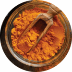
RESEARCH: CURCUMIN ENHANCES LIVER CANCER CHEMOTHERAPY
Liver cancer is incredibly difficult to treat. The cancer cells quickly develop a strong resistance to chemotherapy drugs, including to one of the most commonly used medications, lenvatinib. Fortunately, there is good news. My friend and colleague Dr. Ajay Goel and his team have found curcumin not only helps fight cancer development and growth on its own, but dramatically improves the effectiveness of lenvatinib in stopping aggressive cancer cell growth.
CURCUMIN Enhances Liver Cancer Chemotherapy
Curcumin-Mediated Resistance to Lenvatinib via EGFR Signaling Pathway in Hepatocellular Carcinoma.
Miyazaki K, Morine Y, Xu C, Nakasu C, Wada Y, Teraoku H, Yamada S, Saito Y, Ikemoto T, Shimada M, Goel A. Cells. 2023 Feb 14;12(4):612.
BACKGROUND: Lenvatinib is a multi-kinase inhibitor approved as a first-line treatment for patients with unresectable advanced hepatocellular carcinoma (HCC). However, its response rate is unsatisfactory, primarily due to the acquisition of resistance, which limits its clinical significance for treating patients with HCC. Recent evidence suggests that epidermal growth factor receptor (EGFR) activation can trigger Lenvatinib-resistance; and is considered an important therapeutic target in HCC. Curcumin, one of the most studied naturally occurring botanicals with robust anti-cancer activity, is also reported to be a potent tyrosine kinase inhibitor.
METHODS: In this study, we hypothesized that the anti-EGFR potential of Curcumin might help overcome Lenvatinib resistance in HCC. We established two Lenvatinib-resistant cells and discovered that a combination of Curcumin and Lenvatinib exhibited a synergistic anti-tumor efficacy in the resistant HCC cell lines. In line with previous reports, Lenvatinib-resistant cell lines revealed significant activation of the EGFR, and genome-wide transcriptomic profiling analysis identified that the PI3K-AKT pathway was associated with Lenvatinib resistance.
RESULTS: The combination treatment with curcumin and lenvatinib dramatically suppressed gene and protein expression of the EGFR-PI3K-AKT pathway, suggesting Curcumin overcomes Lenvatinib resistance via inhibition of EGFR. We further validated these findings in tumor spheroids derived from resistant cell lines.
CONCLUSION: In conclusion, we, for the first time, report that Curcumin reverses Lenvatinib resistance in HCC, and that their combination has clinical application potential for adjunctive treatment in HCC.
WHAT THIS MEANS FOR YOUR PATIENTS:
Why Use Curcumin for Liver Cancer?
- Curcumin can stop cancer cells from forming, developing tumors, and spreading
- Curcumin can reduce the toxic effects of cancer drugs
- When used in combination with chemotherapy drugs curcumin has been shown to make them more effective and reduce their toxic effects
Results:
- In tests with lenvatinib-resistant cancer cells, the drug by itself only reduced their number by about 10 to 20%. Curcumin alone did a better job, reducing them by 25 to 50%. But the combination was incredibly effective, reducing cancer cells by almost 75%!
- Cancer cells that were completely resistant to lenvatinib were reduced by 80% when the drug was combined with curcumin
CURCUMIN HELPS DELIVER DRAMATIC RESULTS

Hepatocellular carcinoma (HCC) is the most common form of liver cancer. Diagnosed early, it can be treated with surgery to remove the cancer. However, when surgery is not an option, drugs like lenvatinib are used instead. Unfortunately, levatinib isn’t especially effective (response rates are only about 24 percent), and resistance develops quickly.
Because curcumin can stop cancer cell formation and has a strong track record of enhancing the effects of chemotherapy drugs, researchers tested a specific curcumin extract (BCM95) against liver cancer cells on its own and combined with levatanib. The results were impressive.
Curcumin was incredibly effective against all of the liver cancer cell types used in the study, including drug-resistant cancer cells. But the combination of lenvatinib and curcumin worked best. Together, curcumin and lenvatinib reduced cancer cell colony formation by as much as 72.4%.
One of the reasons HCC develops a resistance to lenvatinib is due to an overexpression of the epidermal growth factor receptor (EGFR) gene, which is also responsible for the survival of cancer stem cells that can cause tumors to return. Understandably, this gene is a frequent target in fighting liver cancer. Fortunately, curcumin is a natural inhibitor of tyrosine kinases, the enzyme family that is home to EGFR. EGFR can act as an “on-switch” for cancer cell growth, so finding that curcumin modulates this activity so powerfully is important to any treatment protocol.
This study represents more evidence to support the addition of curcumin to first-line treatment of cancer, especially for those with limited options to treat their disease. The addition of curcumin could mean the difference between a marginally effective treatment, and a truly life-extending one.
Read More..
Read Less..
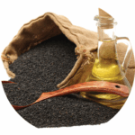
Research: CURCUMIN, BOSWELLIA, & BLACK SESAME SEED OIL DELIVER: 72% PAIN REDUCTION— PAIN RELIEF IN JUST ONE HOUR
Efficacy of high-dissolution turmeric-sesame formulation for pain relief in adult subjects with acute musculoskeletal pain compared to acetaminophen: A randomized controlled study.
Although a lot of attention has been paid to the safety risks and potential for addiction from prescription painkillers, there are plenty of extremely risky over the- counter medicines hidden in plain sight. Acetaminophen (one brand name is Tylenol) is just one of them.
It’s perfectly understandable that when people are in pain, they want relief quickly. But finding an option that is both fast-acting and safe has been nearly impossible—until now. This clinical study on a unique combination of curcumin, boswellia, and black sesame seed oil shows that it reduces pain as quickly and effectively as acetaminophen, but without the risks and dangerous side effects.
Efficacy of high-dissolution turmeric-sesame formulation for pain relief in adult subjects with acute musculoskeletal pain compared to acetaminophen: A randomized controlled study.
RUDRAPPA GH, CHAKRAVARTHI PT, BENNY IR. MEDICINE (BALTIMORE). 2020;99(28):E20373.
BACKGROUND: Acetaminophen (paracetamol) is one of the most commonly used over-the-counter drug for pain relief. Management of acute pain with plant-based nutrients has remained suboptimal due to an absence of data supporting acute relief of pain. In the present study, it was hypothesized that high-dissolution liquid treatment of black sesame extract oil, Curcuma longa and Boswellia serrata may provide pain relief in people with acute musculoskeletal pain as quickly as acetaminophen.
METHODS: In this randomized, active controlled open label study, 88 healthy subjects with acute musculoskeletal pain were randomized to receive treatment capsule (Rhuleave-K; 1,000 mg/d) or 1,000 mg/d acetaminophen for 7 days. Change in pain intensity and pain relief at first 6 hours, 3 days, and 7 days were measured. The onset of analgesia was measured by perceptible pain relief and meaningful pain relief. Other measures were McGill Pain Questionnaire and Patient Global Impression Change.
RESULTS: The treatment formulation resulted in an average magnitude of pain relief comparable to the acetaminophen. 66% of subjects in the treatment group reported a positive response in pain relief (≥50% max TOTPAR; total pain relief) after 6 hours, compared to 73% of control. 73% of subjects on treatment were considered positive responders, compared to 80% in the control group. The average time of onset of analgesia was 1 hour for the treatment group, versus 0.83 hours for the control. At the end of day 3 and 7, there was significant improvement (P<.001 for day 3 and day 7) in the pain condition of the treatment group and was comparable to control (P=.436 for
day 3 and P=.529 for day 7). The total McGill Pain score showed a significant reduction in pain with the treatment irrespective of the pain intensity statistically equal (P=.468) to control. Both the groups were equal in providing sensory pain relief (P=.942), but the treatment was 8.57 times significantly better (P=.027) than acetaminophen in reducing the unpleasantness and emotional aspects (affective domain) involved with acute pain.
CONCLUSION: The results showed that the treatment used in the study may act as a natural, fast-acting, and safe alternative for acute pain relief comparable to acetaminophen.
72% PAIN REDUCTION WHAT THIS MEANS FOR YOUR PATIENTS:
- Average of 72% reduction in pain: equal to acetaminophen, but without harmful side effects
- Perceptible pain relief in the first hour of use
- Works safely and effectively through multiple pain pathways, including COX-2 and 5-LOX
- Can be taken frequently and daily, when needed
- 8.5 times better at reducing emotional distress associated with pain such as feeling tired and fearful
- Safe and effective pain relief without the risk of stroke, heart attack, intestinal bleeding, or damage to liver or stomach
Equally as Fast and Effective as Acetaminophen
This clinical study focused on healthy individuals who suffered from acute pain, much the way anyone does when they have pulled muscles, strained ligaments, or sore joints due to physically demanding jobs, intensive workouts, or some other injury.
The researchers wanted to compare a blend of botanical ingredients including curcumin from turmeric (Curcuma longa), boswellia (Boswellia serrata), and black sesame seed oil to acetaminophen to see how quickly, effectively, and safely the herbal combination could match the frequently used over-the-counter drug.
Curcumin and boswellia have already gained a well-deserved reputation worldwide as extremely effective pain-relievers, fighting COX-2 and 5-LOX inflammation more effectively—and much more safely—than conventional drugs could ever hope to.
The third ingredient in this combination to fight acute pain is black sesame seed oil. It might not seem as familiar in North America, but traditional use and modern research around the world have found black sesame seed oil to be an incredibly valuable nutrient. It provides strong anti-inflammatory power on its own, and like curcumin and boswellia, has a long history of use in Ayurvedic practice.
Most importantly, emulsifying curcumin and boswellia in black sesame seed oil helps these fat-soluble herbs absorb in the intestines and from there, disperse into the bloodstream for optimal pain-fighting ability. This formula creates a powerful concentration of anti-inflammatory ingredients.
On the very first day of the study, the botanical combination virtually matched the perceived pain relief of acetaminophen, with noticeable relief in just one hour. By day seven of the study, it was clear that curcumin, boswellia, and black sesame seed oil were equal to the task of relieving acute pain, reducing it by an average of 72 percent. And the botanical combination was 8.5 times better than acetaminophen at reducing the emotional distress and unpleasantness associated with pain, something that the over-the-counter drug is notoriously unable to do.
There truly is a solution to pain that is safe and effective. This study shows that a combination of curcumin, boswellia, and black sesame seed oil delivers the same relief without the risks of acetaminophen, making it a smart choice for stopping acute pain.
SKIP THE ACETAMINOPHEN:
- According to drugwatch.com, acetaminophen is the leading cause of acute liver failure in the U.S., and accounts for more than 100,000 calls to poison centers.
- Additionally, the use of this readily available over-the-counter drug is responsible for about 60,000 emergency room visits and hundreds of deaths every year in America.
As a healthcare practitioner you are able to offer further guidance on the use of dietary supplements with disease.
Read More..
Read Less..
RESEARCH: RED GINSENG HELPS STOP COLON CANCER
Colon cancer is the second deadliest cancer in America. It is notoriously difficult to treat, because it becomes resistant to chemotherapy drugs, which can be fatal at high dosages. Fortunately, this research shows that red ginseng can be part of a treatment strategy. Researchers found that the botanical switches on naturally protective genes that prevent cancer cells from surviving and tumors from growing. These results are incredibly exciting and offer another desperately needed weapon in the fight against colon cancer.
Ginseng mediates its anti-cancer activity by inhibiting the expression of DNMTs and reactivating methylationsilenced genes in colorectal cancer.
OKUNO K, PRATAMA MY, LI J, TOKUNAGA M, WANG X, KINUGASA Y, GOEL A. CARCINOGENESIS. 2023 MAY 3:BGAD025.
BACKGROUND: Developing safe and effective therapeutic modalities remains a critical challenge for improving the prognosis of patients with colorectal cancer (CRC). In this regard, targeting epigenetic regulation in cancers has recently emerged as a promising therapeutic approach. Since several natural compounds have recently been shown to be important epigenetic modulators, we hypothesized that Ginseng might exert its anti-cancer activity by regulating DNA methylation alterations in CRC.
METHODS: In this study, a series of cell culture studies were conducted, followed by their interrogation in patient-derived 3D organoid models to evaluate Ginseng’s anti-cancer activity in CRC. Genome-wide methylation alterations were interrogated by undertaking MethylationEpic BeadChip microarrays.
RESULTS: First, 50% inhibitory concentrations (IC50) were determined by cell viability assays, and subsequent Ginseng treatment demonstrated a significant anti-cancer effect on clonogenicity and cellular migration in CRC cells. Treatment with Ginseng potentiated cellular apoptosis through regulation of apoptosis-related genes in CRC cells. Furthermore, Ginseng treatment downregulated the expression of DNA methyltransferases (DNMTs) and decreased the global DNA methylation levels in CRC cells. The genome-wide methylation profiling identified Ginseng-induced hypomethylation of transcriptionally silenced tumor suppressor genes. Finally, cell culture-based findings were successfully validated in patient-derived 3D organoids.
CONCLUSION: In conclusion, we demonstrate that Ginseng exerts its anti-tumorigenic potential by regulating cellular apoptosis via the downregulation of DNMTs and reversing the methylation status of transcriptionally silenced genes in CRC.
WHAT THIS MEANS FOR PATIENTS:
- Red ginseng reactivated protective, cancer-stopping genes
- Red ginseng reduced cancer cell colonies by 50%
- High red ginseng dosages dramatically reduced colon cancer cell survival

Anti-Cancer Activity of Red Ginseng
Colorectal cancer is the second leading cause of cancer deaths in the United States. Early screenings beginning at age 50 can help catch pre-cancerous polyps before they develop into tumors, but finding ways of preventing or reducing tumors is still a priority. Fortunately, red ginseng (Panax ginseng) reverses changes that can lead to cancer.
One of the ways the body becomes more susceptible to cancer is when a process called methylation is out of balance and essentially switches off genes in the body that would otherwise stop tumors. The causes behind this can be environmental or dietary reasons, or just a result of getting older. This study found that red ginseng selectively stopped excessive methylation in colorectal cancer (CRC) cells and by doing so, reactivated the natural protective genes that prevent tumor growth. Additionally, it inhibited cancer cell cloning and tumor migration.

The red ginseng (HRG80) used in this study is a source of noble ginsenosides, including Rh2 and Rg3, compounds that have also shown helpful effects on methylation and tumor reduction in breast cancer and ovarian cancer cells in previous research. The concentration of these, and other rare ginsenosides, in this particular red ginseng is due to the plant’s hydroponic cultivation which provides ideal conditions for developing the protective compounds.
These results show that red ginseng can change the course of cancer by changing the epigenetic conditions that could otherwise fuel tumor growth. This should be welcome news to anyone at risk of colorectal cancer looking for safe and effective treatment to tip the balance in their favor.
Read More..
Read Less..